- Visibility 342 Views
- Downloads 46 Downloads
- Permissions
- DOI 10.18231/j.ijos.2020.002
-
CrossMark

A comparative study of the anatomical and functional outcome between Joshi’s External Stabilization System and Kirschner wire fixation technique for Extra-articular Metacarpal fracture
Abstract
Introduction & Objectives: Nowhere in the body, the form and function are so closely related to each other than in hand. Too often these fractures, treated as minor injuries result in major disabilities. Metacarpal fractures comprise between 18–44% of all hand fractures. While many metacarpal fractures do well without surgery, Proper preoperative plannin, surgical intervention wherever needed at a center with backing of equipment and implants with the application of the principles of biological fixation, rigid enough, aids in achieving stable fractures and the ability to allow early mobilization resulting in a good functional outcome. The aim of this study was to compare the outcome between JESS and K wire fixation techniques for extra-articular fractures of the metacarpals.
Materials and Methods: 60 patients were allocated into two groups (Group A - 30 & Group B - 30), Group A underwent K-wire fixation and Group B underwent JESS fixation. Outcomes were measured based on Stability of fixation, tenderness at fracture site, Pin tract infection, Residual fixation, ROM achieved, Wrist and Mayo score, DASH score and VAS scoring system.
Results: Parameters such as the stability of fixation, incidence of pin tract infections, recovery of good range of motion and the outcome was found to be 90%, 20%, 50% (in case of extension) and 83.67% (in case of flexion) and 60%(excellent outcome) respectively in Group B patients who were treated with JESS fixation.
Conclusion: The results of this randomized comparative study showed that JESS fixation was more effective than K-wire fixation in treatment of Extra-articular metacarpal fractures, with emphasis to be provided on regular pin tract dressings and early and effective post-operative mobilization of the joints.
Introduction
Hand is a specialized structure interacting with the environment and is especially sensitive to functional impairment.[1] The human hand has evolved into an organ of exceptional prehensile function, capable of highly complex movements and manipulation. Hand injury is extremely common and accounts for about 15% of the casualities presenting to the emergency department.[2]
Hand is more prone to variety of fractures by multiple causes. Some of the most common causes of hand injuries are crush/compression injuries, blunt trauma, fall, road traffic accidents, machinery injury, sports related activity, explosions and firearm injuries.[4], [3] Hand fracture especially to metacarpals and phalanges are the most common and are often neglected as minor injuries which accounts for 15% of the admissions to the emergency departments.[5] The incidence is more at the age of 10–40 years with male dominanance.[6]
The metacarpal fractures comprise between 18–44% of all hand fractures. Non thumb metacarpals account for around 88% of all metacarpal fractures, with the fifth finger most commonly involved. Metacarpal and phalanx fractures comprise 46% of the hand fractures and out of that mostly involved are the proximal phalanx and metacarpal neck fractures are most common and then the middle and distal phalanx and the base of metacarpal. The majority of the metacarpal fractures are isolated injuries, simple, closed and stable.[7]
The fractures of the hand cause significant disability and may lead to long term negative functional sequelae, such as the loss of ability to work and live at the preinjury level.
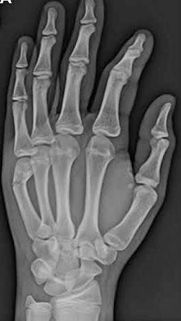
A variety of methods and algorithms are available to treat the metacarpal fractures. Various modes of treatment have been used for fractures of the hand which include K-wire fixation, mini plates, external fixator application systems.
Surgical intervention wherever needed, application of the principles of fixation, a system rigid enough to allow early mobilization are all very important for a good functional outcome.[8]
The treatment of hand deformities involves development of muscle power, increase in joint range and redevelopment of coordination. Conservative method is the primary treatment of choice but fixation is need in few unstable fracture, multiple fractures, intra-articular fractures and open fractures to obtain the optimal position for bone healing and to allow early movement.[9]
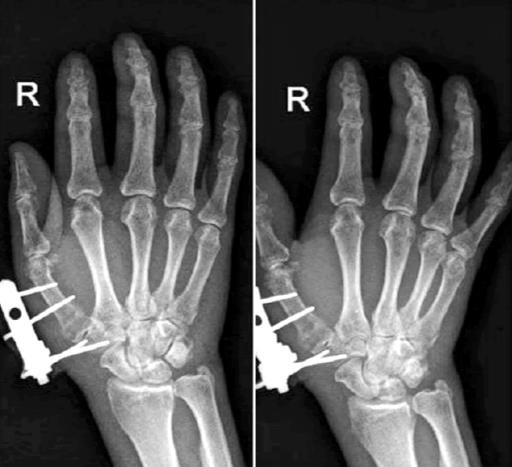
External stabilization system is an effective treatment modality for unstable and compound injuries. With the use of thin smooth wires, which are places away from the injury site in a stable configuration, Joshi’s external stabilizing system (JESS) provides a stable skeletal environment aiding rapid healing of soft tissue wit h establishment of microvascular circulation, immediate active and passive mobilization of the uninjured adjacent joint.[10]
JESS is a simple, versatile and light weight fixation with the added possibility of incorporation of splints or conversion to dynamic mobilization units. JESS provides rigid fixation of bones in which other forms of immobilization are not appropriate e.g., open fracture. It is possible to compress, neutralize or distract a fractures fragment and also allowing aggressive and simultaneous treatment of bone and soft tissue lesions. It is possible to immediately move the proximal and the distal joints, thereby reducing edema, preventing capsular fibrosis, joint stiffness and muscularatrophy.[11]
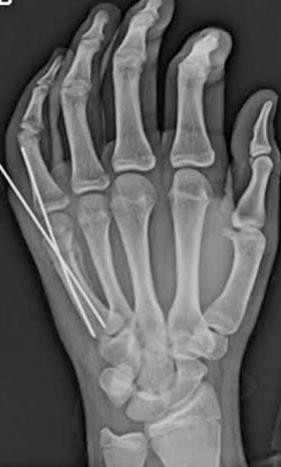
Kirschner- wires are most versatile, simplest and cheapest method of fixing hand injuries. They can be introduced percutaneously without exposing the fractures. It is sufficiently stable to allow early motion without subjecting the hand to surgical trauma of openreduction. [11]
Methodology
A hospital based prospective clinical study was undertaken in the Department of Orthopaedics of Rajarajeswari Medical College & Hospital, Bengaluru among the patients admitted for unstable extra-articular fractures of the metacarpals. A total of 60 patients with metacarpal fractures constituted the study sample. Clearance from the institutional ethics committee was obtained before the study was started. A bilingual, written and informed consent was obtained from all the cases before they were included in to the study. The inclusion and exclusion criteria were as follows,
Inclusion criteria
Skeletally mature patients (20-60 years)
Closed and Open fractures of metacarpals (Type 1, 2 and 3A only).
Extra-articular fractures of metacarpals.
Physically fit for surgery
Patient consent.
Exclusion criteria
Intra-articular fractures.
Crush injuries of the hand with multiple compound Grade 3 fracture.
Associated co-morbidities.
Infection at site of procedure.
Patient refusal.
A total of 60 subjects were considered for the study, with them being divided equally into two groups
The percutaneous intramedullary Kirschner -wire fixation group (Group A).
Joshi’s external stabilization system fixation group (Group B).
The injuries were classified on the basis of fracture level and type. Assessment of patients was carried out according to the Mayo Wrist and DASH Scoring systems. Finally, Radiographic and clinical outcomes of both groups was assessed and compared.
A detailed history taken before the patients were subjected for the surgery. The patients were also subjected for the detailed physical examination. X-ray with AP, oblique/lateral views were done, Routine investigations as per the institution protocol were sent. All the patients were subjected for Pre-anesthetic check-up and clearance. In case of open fractures, debridement of the wound and thorough irrigation was done with normal saline.
Closed/open reduction was achieved by traction and manipulation. To maintain reduction, percutaneous Kirschner-wire or Joshi’s external stabilization system were used. Image intensifier (C-arm) was used as a guide for the steps mentioned above. Post-operatively, x-rays were taken to evaluate the fixation. Patients were taught active mobilization of the unaffected fingers, elbow and shoulder from immediate post-op period. Pin tract dressings were done regularly. Patients were called for periodic evaluation at 2 weeks, 4 weeks, 6 weeks and 8 weeks on OPD basis to assess:
Stability of fixation.
Tenderness at fracture site.
Pin tract infections.
Residual stiffness.
Joshi’s external stabilization system or Kirschner-wire removal was done at 3 to 6 weeks interval with immediate vigorous mobilization of the immobilized joint to avoid stiffness. Functional outcome was assessed based on the total active range of movement in degrees of each injured finger separately.
The data was obtained by using Predesigned and pretested proforma. The data was entered in Microsoft excel sheet and later transferred and analyzed using Statistical Package for Social Services (SPSS vs 20). The qualitative data was presented as frequencies and percentages and quantitative data was presented as mean and standard deviations. Chi square test was used as test of significance for the qualitative data.
Results
In our study in Group A, no patients below 20 years of age were operated. 16.7% of the patients were in the age group of 21-30 years. 23.3% of the patients were in between 31-40 years. Most of the patients in Group A belonged to 41 – 50 years. In the group B, 33.3% of the patients belonged to 21–30 years of age. 16.7% belonged to age group of 31-40 years. 10% belonged to age group of 41-50 years. 20% belonged to age group of 51-60 years and finally, 13.3% belonged to the age group of more than 60 years. This difference in age was not statistically significant between the two groups.
In Group A, 56.7% of the patients were found to be males in comparison to 43.3% of the patients being females. In Group B, an equal incidence of 50% on each side was documented. There was no statistically significant difference in sex between the two groups.
About 40% of the patients in Group A and 43.3% of the patients in Group B had the injury due to road traffic accidents. 16.7% in Group A and 20% of patients in Group B were found to have sustained the injury due to an agriculture related injury. 20% in Group A and 33.3% of the patients in group B sustained the injury due to industrial accidents. Finally, 23.3% belonging to group A and only 3.3% of the patients belonging to group B sustained the injury due to a road traffic accident. This difference in mode of injury was not statistically significant between the two groups.
About 43.3% of the patients in Group A were drivers and 33.3% of the patients in Group B were industrial workers. This difference in occupation was not statistically significant.
An equal incidence of 50% on right and left side was found in patients belonging to group A. The right hand was injured in 63.3% of the patients in Group B as compared to 36.7% of affection to the left hand. This difference in the side affected was not statistically significant.
First and second metacarpal was affected equally in 33.3% of the group A patients, 10% of patients had a third metacarpal injury, 6.7% of the patients sustained injury to the fourth metacarpal and 16.7% of the patients had an injury to the fifth metacarpal. About 23.3% of the patients had first and third metacarpal injury in case of the group B, 20% in case of the second and fifth metacarpal and finally, 13.3% of the patients were found to have sustained injury to the fourth metacarpal. This difference in metacarpal involved was not statistically significant.
About 63.3% of patients and 13.3% of patients of group A and group B respectively had Type I fractures. 30% of group A and 50% of the patients of group B had Type II fractures. 6.7% of group A and 36.7% patients of group B had Type IIIA fractures. This difference in type of the fractures was statistically significant between the two groups.
Mean time to surgery was 2.8 days in group A and 3.03 days in group B which was not statistically significant.
The mean healing time in Group A was 7 weeks and in group B was 8.2 weeks, which was statistically significant between the two groups.
Only 66.7% of the patients in group A had stable fixation as compared to 90% of the patients in group B. This difference in stability of the fixation was statistically significant.
Tenderness at the fracture site was present in 56.7% of the group A patients as compared to only 26.7% of the group B patients. This difference in tenderness was statistically significant.
The pin tract infection was present in 46.7% o f the group A patients as compared to 20% of the group B patients. This difference in pin tract infection was statistically significant between the two groups.
The residual fixation was done in 23.3% of the cases in group A and 13.3% of the patients in group B. This difference in residual fixation was not statistically significant.
50% of the patients of group B were found to have complete extension at the MCP joint in comparison to 13.3% of the group A patients where complete extension was achieved. 3.3% of the patients of group A were found to have a 5° extension lag as compared to 13.3% in group B. The extension lag was found to be 10° for 20% of group A and 13.3% of group B patients. Most patients of group A were found to have an extension lag of 20° in group A as compared to 13.3% patients of group B. An equal incidence of 10% was found between the two groups for an extension lag of 30°. Further, 13.3%, 6.7% and 10% of extension lag of 40°, 50° and 60° respectively was found in patients of group A. This difference in extension lag was statistically significant between the two groups.
The mean flexion in Group A patients was 75.5 degrees and group B patients was 83.67 degrees. This difference in flexion was statistically significant between the two groups.
The mean Mayo and wrist score in group A was 72.33 and group B was 79. This difference in Wrist and mayo score was statistically significant between the two groups.
The mean Dash score in group A was 34 and group B was 20. This difference in DASH score was statistically significant between the two groups.
The mean VAS score in group A was 3.97 and group B was 2.93. This difference in mean VAS scores was statistically significant between the two groups.
Only 26.7% of the patients in group A as compared to 60% in group B had excellent results. The study revealed a poor outcome in 13.3% of the group A and 10% of the group B patients. This difference in outcome was statistically significant between the two groups.
| Group A | Group B | ||
| n (%) | n (%) | ||
| Age in Years | ≤ 20 | 0 | 2 (6.7) |
| 2 1- 30 | 5 (16.7) | 10 (33.3) | |
| 31 – 40 | 7 (23.3) | 5 (16.7) | |
| 41 – 50 | 11 (36.7) | 3 (10.0) | |
| 51 – 60 | 5 (16.7) | 6 (20.0) | |
| > 60 | 2 (6.7) | 4 (13.3) | |
| Total | 30 (100) | 30 (100) | |
| Sex | Male | 17 (56.7) | 15 (50.0) |
| Female | 13 (43.3) | 15 (50.0) | |
| Total | 30 (100) | 30 (100) | |
| Mode of Injury | Agriculture | 5 (16.7) | 6 (20.0) |
| Industrial | 6 (20.0) | 10 (33.3) | |
| RTA | 12 (40.0) | 13 (43.3) | |
| Trauma | 7 (23.3) | 1 (3.3) | |
| Total | 30 (100) | 30 (100) | |
| Occupation | Business | 9 (30.0) | 4 (13.3) |
| Driver | 13 (43.3) | 4 (13.3) | |
| Farmer | 5 (16.7) | 6 (20.0) | |
| Industrial worker | 6 (20.0) | 10 (33.3) | |
| IT worker | 3 (10.0) | 3 (10.0) | |
| Student | 2 (6.7) | 3 (10.0) | |
| Total | 30 (100) | 30 (100) | |
| Side of the hand | Left | 15 (50.0) | 11 (36.7) |
| Right | 15 (50.0) | 19 (63.3) | |
| Total | 30 (100) | 30 (100) | |
| Metacarpal Involved | First | 10 (33.3) | 7 (23.3) |
| Second | 10 (33.3) | 6 (20.0) | |
| Third | 3 (10.0) | 7 (23.3) | |
| Fourth | 2 (6.7) | 4 (13.3) | |
| Fifth | 5 (16.7) | 6 (20.0) | |
| Total | 30 (100) | 30 (100) | |
| Type of fracture | Type I | 19 (63.3) | 4 (13.3) |
| Type II | 9 (30.0) | 15 (50.0) | |
| Type IIIA | 2 (6.7) | 11 (36.7) | |
| Total | 30 (100) | 30 (100) |
| Group A | Group B | ||
| Time of surgery (days) | Mean ± SD | 2.8 ± 1.03 | 3.03 ± 1.03 |
| Healing time (in weeks) | Mean ± SD | 7.0 ± 1.29 | 8.2 ± 1.67 |
| Stability of fixation | Stable | 20 (66.7) | 27 (90.0) |
| Unstable | 10 (33.3) | 3 (10.0) | |
| Total | 30 (100) | 30 (100) | |
| Tenderness | Absent | 13 (43.3) | 22 (73.3) |
| Present | 17 (56.7) | 8 (26.7) | |
| Total | 30 (100) | 30 (100) | |
| Pin Tract Infection | Absent | 16 (53.3) | 24 (80.0) |
| Present | 14 (46.7) | 6 (20.0) | |
| Total | 30 (100) | 30 (100) | |
| Residual Fixation | Not done | 23 (76.7) | 26 (86.7) |
| Done | 7 (23.3) | 4 (13.3) | |
| Total | 30 (100) | 30 (100) | |
| Extension lag (in degrees) | 0 | 4 (13.3) | 15 (50.0) |
| 5 | 1 (3.3) | 5 (13.3) | |
| 10 | 6 (20.0) | 4 (13.3) | |
| 20 | 7 (23.3) | 4 (13.3) | |
| 30 | 3 (10.0) | 3 (10.0) | |
| 40 | 4 (13.3) | 0 | |
| 50 | 2 (6.7) | 0 | |
| 60 | 3 (10.0) | 0 | |
| Total | 30 (100) | 30 (100) | |
| Flexion in Degrees | Mean ± SD | 75.5 ± 15.11 | 83.67 ± 8.09 |
| Wrist and Mayo score | Mean ± SD | 72.33 ± 12.23 | 79.0 ± 8.85 |
| DASH Score | Mean ± SD | 34.0 ± 24.3 | 20.0 ± 15.76 |
| VAS Score | Mean ± SD | 3.97 ± 1.69 | 2.93 ± 1.23 |
| Outcome | Excellent | 8 (26.7) | 18 (60.0) |
| Good | 7 (23.3) | 6 (20.0) | |
| Satisfactory | 4 (13.3) | 3 (10.0) | |
| Poor | 4 (13.3) | 3 (10.0) | |
| Total | 30 (100) | 30 (100) |
Discussion
This study was mainly undertaken to compare and report the outcome of unstable extra-articular metacarpal fractures treated using intramedullary Kirschner-wires and Joshi’s external fixator system and to determinate which of the two techniques provides better clinical and radiographic results.
Age group
Most of the patients in Group A were aged between 41–50 years and 21–30 years of age in groups B. In a study by Naidu et al, the incidence of fractures were more in the age group of 31 – 40 years in males and 21 – 30 years and more than 51 years in females.[10] A study by Rajkumar et al had reported that, metacarpal fractures were most common in the age less than 30 years.[11] A study by Salunkhe et al had reported that the mean age was 30 years.[12]
Sex
Male patients outnumbered female patients in this study. A study Naidu et al also observed similar findings.[10] A study by Rajkumar et al had reported that males were commonly affected than females.[11] Salunkhe et al had also reported similar findings.[12]
Road traffic accidents
About 40% of the patients in Group A and 43.3% of the patients in Group B had the injury due to road traffic accidents. Naidu et al reported that, injury by road traffic accidents and injury by machinery were common mode of injuries.[10] Rajkumar et al also reported the same followed by assault injuries and violence.[11]
Occupation
About 43.3% of the patients in Group A were drivers and 33.3% of the patients in Group B were industrial workers. No studies compared these results.
Side affected
Right hand was affected in 50% of the group A and 63.3% of the patients in Group B. A study by Rajkumar et al had reported that right side was affected more than the left side.[11] Salunkhe et al had reported no association between the right or left side.[12]
Metacarpal affected
First and second metacarpal was affected in 33.3% of the group A patients. About 23.3% of the patients had first and third metacarpal injury. No studies had reported the findings of this study.
Type of fracture
This study had shown that, about 63.3% of the patients had Type I fractures and 50% of the patients had Type II fractures. A study by Naidu et al had reported grade 3 injuries more than other type of injuries.[10]
Time to surgery
Mean time to surgery was 2.8 days in group A and 3.03 days in group B. No studies have reported such results.
Healing time
The mean healing time in Group A was 7 weeks and in group B was 8.2 weeks which was significantly more in Group B. A study by Naidu et al had reported that non-union was common in 8% and delayed union was reported in 10% of the cases. Post-operative wounds healed within 4 weeks in 84% of the cases and in 26% of the cases it was observed to heal within 8 weeks.[10] A study by Gupta et al had reported that the fracture healing time was between 8 – 12 weeks in more than half of the cases.[13]
Stability of fixation
About 66.7% of the patients in group A had stable fixation and 90% of the patients in group B had stable fixation which was statistically significant. Naidu et al had reported loosening of K wires in 16% of the cases.[10]
Tenderness
Tenderness was present in 56.7% of the group A patients and 26.7% of the group B patients which was statistically significant. A study by Naidu et al had reported swelling and tenderness as the most common post-operative complication in 32% of the cases.[10]
Pin tract infection
The pin tract infection was present in 46.7% of the group A patients and 20% of the group B patients significantly higher in Group A. A study by Naidu et al had reported pin tract infections in 14% of the cases.[10] A study by Gupta et al had reported pin tract infection in 12 patients.[13]
Residual fixation
The residual fixation was done in 23.3% of the cases in group A and 13.3% of the patients in group B. Studies were not available to compare these findings.
Extension lag
The extension lag in 23.3% of the patients in group A was 20 degrees and 0 degree in 50% of the cases of group B which was statistically significant. No studies compared these results.
Flexion
The mean flexion in Group A patients was 75.5 degrees and group B patients was 83.67 degrees was significantly more in group B. Studies were not available to compare these findings.
Wrist and Mayo score
The mean mayo score in group A was 72.33 degrees and group B was 79 which significantly higher in group B. No studies reported these findings.
DASH score
The mean Dash score in group A was 34 and group B was 20 and was significantly lower in Group B. The studies were not available to compare these findings.
VAS scores
The mean VAS score in group A was 3.97 and group B was 2.93. This difference in mean VAS scores was statistically significant between the two groups. None of the studies were available to compare these findings.
Outcome of the surgery
About 26.7% of the patients in group A and 60% in group B had excellent results. This difference in outcome was statistically significant between the two groups. A study by Rajkumar et al reported that, excellent results were observed in 57.7% of the JESS method and 34.1% of the internal fixation method.[11] A study by Salunkhe et al had reported excellent outcome in 24 cases, 5 cases had good outcome and only 1 case had satisfactory outcome.[12] In a study conducted by Gupta et al, good outcome was significantly associated with the younger age group in 37.77% of the cases.[13]
Conclusion
Our study showed that JESS group had shown longer time of healing, more stable fixation, less number of complications, less number of repeated surgeries, more range of mobility of the fracture site, higher Wrist and Mayo scores, Lower DASH and VAS scores, excellent to good outcome. The procedure is simple, easy to perform and requires less time and expertise. Hence, the study concludes that JESS fixation was superior than K wire fixation. But this study is without limitations. The sample size was not calculated in this study. The scanty literature preambles the need for more studies like this to bring out more facts about the use of JESS fixation in metacarpal injuries.
Source of funding
None.
Conflict of interest
None.
References
- Diaz-Garcia R, Waljee JF. Current Management of Metacarpal Fractures. Hand Clinics. 2013;29(4):507-518. [Google Scholar]
- Crockett D. Rigid fixation of bones of the hand using K wires bonded with acrylic resin. Hand. 1974;6(1):106-107. [Google Scholar]
- Fitoussi F, IP WY, Chow SP. External fixation for comminuted phalangeal fractures a biomechanical cadaver study. J Hand Surg Br. 1996;21(6):760-764. [Google Scholar]
- Joshi BB. Percutaneous Internal Fixation of Fractures of the Proximal Phalanges. Hand. 1976;8(1):86-92. [Google Scholar]
- Stern PJ, GD, HR, PW. Factures of metacarpals and phalanges. Green’s operative hand surgery. 4th edn. 1999;1:711-757. [Google Scholar]
- Jonge Jd, Kingma J, Lei Bvd, Klasen H. Fractures of the metacarpals. A retrospective analysis of incidence and aetiology and a review of the English-language literature. Inj. 1994;25(6):365-369. [Google Scholar]
- Gudmundsen TE, Borgen L. Fractures of the fifth metacarpal. Acta Radiol. 2009;50(3):296-300. [Google Scholar]
- Schuind F, Donkerwolcke M, Burny F. External Minifixation for Treatment of Closed Fractures of the Metacarpal Bones. J Ortho Traumat . 1991;5(2):146-152. [Google Scholar]
- Freeland AE, Orbay JL. Extra-articular Hand Fractures in Adults. Clin Orthop Relat Res. 2006;PAP:133-145. [Google Scholar]
- Naidu KVD. Management of metacarpal and phalangeal fractures with JESS fixator: A prospective study. IJOS. 2018;4:383-387. [Google Scholar]
- Rajkumar K, Azeem MA. Comparison of external and internal fixation methods for the metacarpal fractures. IJOS. 2016;2:322-325. [Google Scholar]
- Salunkhe RM, Joshi H, Pisal TK, Biswas SK, Patel JJ, Singh A. A prospective study of dynamic treatment of fracture phalanx and metacarpals of the hand with Kirschner-wire fixation/external fixator and finger splint: Daycare management (30 cases). Med J DY Patil Univ . 2016;9(6):708-713. [Google Scholar]
- Gupta AK, Sapra R, Kumar R, Gupta SP, Kaushik D, Gaba S, et al. Role of Joshi's external stabilization system with percutaneous screw fixation in high-energy tibial condylar fractures associated with severe soft tissue injuries. Chin J Traumatol. 2015;18(6):326-331. [Google Scholar]
- Abstract
- Introduction
- Methodology
- Results
- Discussion
- Age group
- Sex
- Road traffic accidents
- Occupation
- Side affected
- Metacarpal affected
- Type of fracture
- Time to surgery
- Healing time
- Stability of fixation
- Tenderness
- Pin tract infection
- Residual fixation
- Extension lag
- Flexion
- Wrist and Mayo score
- DASH score
- VAS scores
- Outcome of the surgery
- Conclusion
- Source of funding
- Conflict of interest
- References