- Visibility 118 Views
- Downloads 38 Downloads
- DOI 10.18231/j.ijos.2020.019
-
CrossMark

Functional outcome of submuscular plating for diaphyseal long bone fractures
Introduction
Incidence of humerus shaft fracture in adult population is 3 to 5% approximately and it comprises 20% of all humerus fractures.[1], [2] Majority of these fractures can be treated by conservative management using U shaped cast, velpau sling, thoracobrachial cast, brachial orthosis.[3], [4], [5] However this can lead to nonunion, delayed union, malnuion, restricted elbow and shoulder movements.[4], [6] The surgical treatment includes either open reduction and internal fixation with plating or closed reduction and internal fixation with nailing.[7], [8], [9] Intramedullary nailing is generally preferred in comminuted, compound and pathological fracture whereas plating is preferred in fracture with minimal comminution and the one which requires exploration of the radial nerve.[10], [11] Open reduction and internal fixation helps to achieve anatomical reduction but this technique requires longer surgical duration, large incision, more soft tissue dissection, blood loss and periosteal stripping which can lead to increased chances of nonunion, infection and wound healing problems.[12] The incidence of nonunion with such techniques is 2 to 10%, infection 2 to 4% and radial nerve palsy is 2 to 5%.[9] Femoral shaft fractures account for 1.4% to 1.7% of all fractures seen in paediatric population.[10] Treatment for paediatric femoral fractures has been evolving from conservative to operative intervention especially in older children, more than 5 years of age.
Children younger than 6 years of age can be managed conservatively by traction and spica cast application.[11] Increased cost of hospitalization, probability of malalignment and prolonge dimmobilization due to conservative method has led to shift towards operative intervention in older children. Different modalities described are intramedullary nailing by flexible or rigid nails, external fixation, traditional open reduction and plate fixation and sub muscular bridge plating. Goals of any operative treatment modality are to preserve femoral blood supply, avoid damage to the physis and achieve adequate fracture stability. External fixation may lead to refracture, malunion, delayed union, pin tract infections and unsightly scars.[12], [13], [14], [15] Rigid intramedullary nails with piriformis fossa as the entry site raise possibility of damaging the vascular supply to the femoral head, resulting in avascular necrosis. Utilizing nails with greater trochanter entry points also do not completely obviate this problem.
Fractures in proximal 1/3rd and distal 1/3rd may not gain adequate stability with intramedullary nails due to smaller length of nail-bone contact.[16], [17], [18], [19] Traditional open reduction and application of compression plate requires long incisions and more soft tissue dissection. Therefore it has a higher risk of infection and delayed healing with a reported reoperation rate of 10%.[20]
Sub-muscular plating for diaphyseal long bone fractures was first reported for adult patients in the late 1990s.[21] As the procedure began to gain acceptance among Orthopaedic surgeons similar principles were applied for treatment of fractures in the pediatric population also. The advantages include a minimally invasive, soft tissue preserving approach, relative stability that allows for early ROM and reliable healing. In this study we have evaluated the results of submuscular plating in diaphyseal long bone fractures.
Materials and Methods
We evaluated the clinical, radiological and functional results of Submuscular plating in 30 patients operated in between June 2016 to June 2019 for fracture shaft humerus, shaft of femur and shaft of tibia. All the surgeries were carried out by a single surgical team at a single institute. The inclusion criteria for the study were: Fracture shaft of humerus, shaft of femur, shaft of tibia; Fractures without any neurological deficit; Patients with minimum 2 year follow up.
The exclusion criteria for the study were: Compound fractures; Fractures with non union or delayed union; Pathological fractures; Neurovascular insufficiency.
Operative technique for humerus
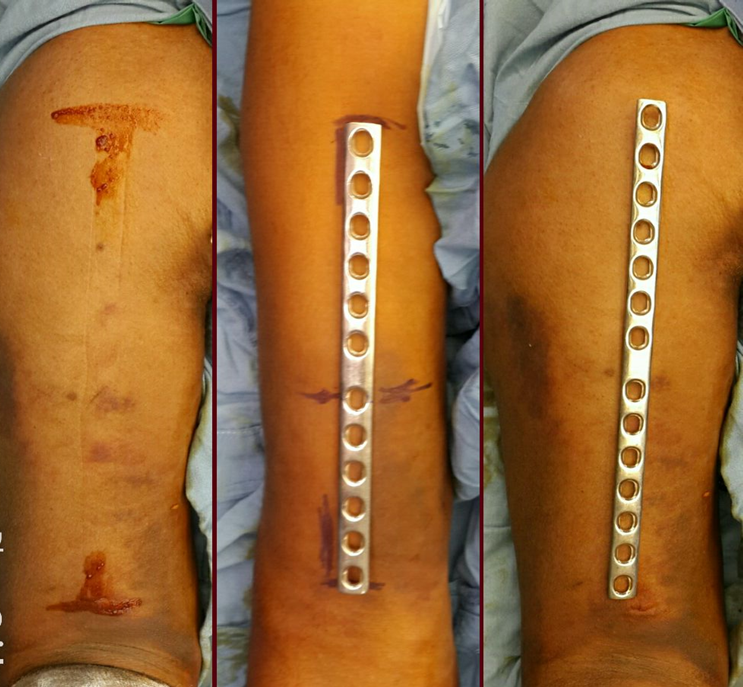
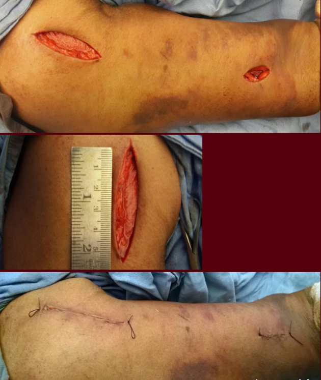
The surgery was carried out in a beach chair position with the arm abducted about 400 – 600 and supine under general anaesthesia. Indirect fracture reduction was achieved manually. With the help of C arm length of the plate, proximal and distal screw placement and skin incision was determined by keeping plate on the skin anteriorly 4-5 cm incision was made distally along lateral border of biceps approximately 5 cm proximal to flexion crease. After this an interval was made between the biceps tendon and brachioradialis muscle to expose brachailis. By blunt dissection an interval was made in the fibers of brachialis till the anterior surface of humerus was seen. Then 4-5 cm incision was made proximally and an interval was made between lateal border of proximal biceps and medial border of deltoid. An epiperiosteal tunnel connecting the two incisions was made using a plate itself. From distal to proximal incision longest possible predermined 4.5mm narrow DCP or LCDCP was slide in the tunnel. Contouring of the plate was not essential as the implant was used to provide indirect relative stable fixation and minimal cortical contact preserving periosteal blood supply.[22] Under C arm control traction was applied to restore length and any angular or rotational deformity was corrected manually. Where reduction was difficult best possible reduction was accepted. After ensuring that plate is positioned centrally on anterior surface and reduction is satisfactory it was fixed with 2 screw on each side in most proximal and most distal holes of the plate. While putting screw reduction was held by assistant and repeatedly checked under C arm. The wound was closed in layers and sterile dressing applied. The operative time was recorded from incision to closure of wound. The arm was immobilized in a cuff and collar sling
Post operatively adequate antibiotic cover was given. Active shoulder and elbow exercises within pain limits were started on 2nd post op day. Patients were discharged on 5th post op day. Patients were followed up periodically till radiological bony union occurred and half yearly thereafter. Radiological assessment was done on standard anteroposterior and lateral view. At every follow up, each patient was evaluated clinically, radiographically and functionally for the signs of union, nonunion, malunion, infection.
Operative technique for femur
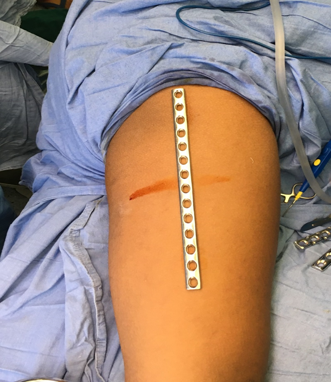
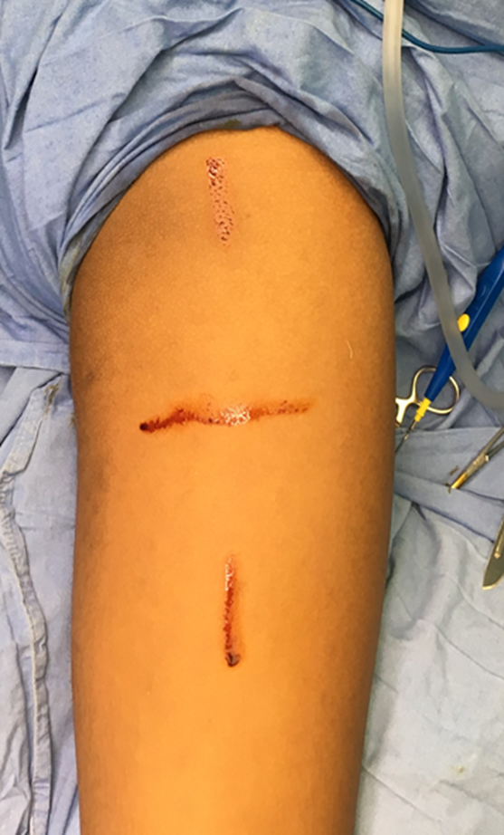
Patient supine on operation table, under all aseptic precautions scrubbing, painting and draping done. Depending on fracture site, proximal incision (4-6 centimeters long) was usually made at the level of the vastus ridge on the greater trochanter. Dissection was done to identify plane between muscle mass and periosteum over lateral cortex of femur and this plane was developed distally using a long Cobb’s elevator. 4.5mm narrow low contact dynamic compression plate (LC-DCP) plates were utilized The plate was slide in this plane from proximal to distal staying epi-periosteal. Position of plate was provisionally secured with a 1.5mm K-wire through the plate hole at one end, utilizing intraoperative imaging. The position of the other end of the plate was determined under fluoroscopy and incision was made at that level. Distal incisions were made first when fracture was in distal half of bone and plate was slide from distal to proximal in similar epiperiosteal manner. Fracture was reduced with manipulation and longitudinal traction. We used folded sterile sheets as adjunct for reduction whenever necessary. If reduction was acceptable reduction position of plate was adjusted to maintain plate in good contact with bone and 2nd K-wire was introduced through a hole at the other end of the plate for provisional fixation. After additional evaluation and necessary adjustments were made, 3 cortical screws were inserted in either fragments. In few cases soft tissue interposition made closed reduction difficult and an incision was made at fracture site to achieve reduction with finger manipulation or a bone hook. We did not use any splints in postoperative period.
Postoperatively we mobilized the patients within 1-3 days as per their comfort, using a walker and with the recommendation to be partial weight bearing for 6 weeks.
Operative technique for tibia
The surgery was carried out in supine position with angle frame under spinal anaesthesia/general anaesthesia. All surgeries were carried out by single set of surgeons. Indirect reduction was achieved manually. With the help of C arm length of plate, proximal and distal screw placement and skin incision was determined by keeping plate on anterolateral aspect of proximal tibia. Anterolateral approach used for the exposure.4-5 cm Straight incision lateral to patella taken till tibial tuberosity. Deep fascia anterior to the IT band exposed, Proximal attachment of Tibialis anterior muscle released, anterior tunnel made in the submuscular plane, longest possible 4.5 mm LCDCP/DCP slide from proximal fragment to distal fragment. Reduction achieved with controlled traction under C arm guidance, An epiperiosteal tunnel connecting the two incisions was made, precontured plate was slide in the tunnel, three proximal and three distal screws are placed, the wound was closed in layers, sterile dressing done. Bed side knee, hip, ankle were started on 1 st post op day or as per patients comfort, patient was discharged on 5th post op day, patients was mobilized with walker with non weight bearing for 6 weeks, partial weight bearing for next 6 weeks and full weight bearing after 12 weeks.
Results
The study included 17 male and 13 female patients with mean age of 28 years (range 10-50 years). All patients were operated within 5 days after injury. AO-ASIF Trauma classification was used to classify the fracture. Mechanism of injury was high velocity injury in 18 with 08 polytrauma patients and domestic fall in 12. The mean surgical time was 88 minutes (62-140 min). All fractures united with mean time of 16 weeks without any further intervention. (Femur takes more time to unite than humerus). None of the patients had any major complication except superficial infection in two. The mean surgical time was 88 minutes (range 62-140). 12 to 15 hole plate were used commonly. Average blood loss was 100 cc.
| S.No. | Age | Sex | Fractured bone | Associated Injury | SurgicalTime(min) | Follow up(wks) | Union (wks) | Implant type | Complication |
| 1 | 10 | M | Femur | 120 | 30 | 20 | 4.5DCP | ||
| 2 | 38 | M | Tibia | 80 | 28 | 16 | 4.5LCDCP | ||
| 3 | 12 | F | Femur | RADIUS# | 100 | 30 | 18 | 4.5DCP | |
| 4 | 28 | M | Femur | 105 | 28 | 16 | 4.5DCP | ||
| 5 | 40 | F | Humerus | 76 | 26 | 14 | 4.5LCDCP | Infection | |
| 6 | 38 | F | Humerus | 88 | 22 | 16 | 4.5LCDCP | ||
| 7 | 22 | M | Tibia | NOF# | 70 | 30 | 18 | 4.5LCDCP | |
| 8 | 15 | F | Femur | 98 | 28 | 14 | 4.5DCP | ||
| 9 | 36 | F | Humerus | TIBIA# | 80 | 30 | 13 | 4.5LCDCP | |
| 10 | 12 | F | Tibia | 75 | 28 | 16 | 4.5LCDCP | ||
| 11 | 27 | M | Tibia | 62 | 30 | 16 | 4.5LCDCP | ||
| 12 | 45 | M | Humerus | 79 | 26 | 13 | 4.5LCDCP | ||
| 13 | 17 | M | Femur | CLAVICLE# | 140 | 30 | 17 | 4.5DCP | |
| 14 | 35 | M | Tibia | 90 | 30 | 18 | 4.5LCDCP | ||
| 15 | 30 | F | Humerus | 82 | 28 | 15 | 4.5DCP | ||
| 16 | 30 | M | Femur | 110 | 30 | 20 | 4.5DCP | ||
| 17 | 35 | M | Tibia | 81 | 28 | 16 | 4.5LCDCP | ||
| 18 | 12 | F | Femur | HUMERUS# | 90 | 30 | 18 | 4.5DCP | |
| 19 | 17 | M | Femur | 95 | 28 | 18 | 4.5DCP | ||
| 20 | 45 | F | Humerus | 76 | 26 | 14 | 4.5LCDCP | ||
| 21 | 27 | F | Humerus | 78 | 22 | 16 | 4.5LCDCP | ||
| 22 | 16 | M | Tibia | ULNA# | 74 | 30 | 18 | 4.5LCDCP | |
| 23 | 36 | F | Femur | 98 | 28 | 17 | 4.5DCP | Infection | |
| 24 | 40 | F | Humerus | TIBIA# | 86 | 30 | 13 | 4.5LCDCP | |
| 25 | 22 | F | Tibia | 75 | 28 | 16 | 4.5LCDCP | ||
| 26 | 36 | M | Tibia | 62 | 30 | 16 | 4.5LCDCP | ||
| 27 | 38 | M | Humerus | 79 | 26 | 13 | 4.5LCDCP | ||
| 28 | 50 | M | Femur | BOXER’S# | 130 | 30 | 17 | 4.5DCP | |
| 29 | 30 | M | Tibia | 90 | 30 | 16 | 4.5LCDCP | ||
| 30 | 28 | M | Humerus | 82 | 28 | 15 | 4.5DCP |


Discussion
Minimally invasive technique achieves relative stability by indirect reduction. It is routinely used for treating fractures of tibia and femur since 1980's, but use of this technique for treatment of humerus fracture has been described recently.[23], [24] Submuscular plating for diaphyseal long bone fracture is basically a MIPO technique. In our series plate fixation was carried out with only 2 to 3 screws, far from fracture site on each side, using longest possible plate. Using a long plate improves the lever arm of the construct which leads to a low pull out force acting on each screw.[25] The construct can withstand considerable deformation forces as bending stresses are distributed over a long segment of the plate, reducing the chances of plate failure.[26] In our study we have used C arm for assessing and holding the reduction. We operated all the cases within 5 days after injury as reduction becomes difficult by indirect measure in older fractures.
The fracture fixation which allows the micro movements at the fracture site under physiological stress are called as flexible fixations which aids in early union by callus formation. The healing by bridging callus is faster, effective and has more strength as compared to primary bony healing.[26] The primary bone healing without callus formation is not very strong and has risk of refracture after removal of implant which happens in the open technique.[27] It preserves blood supply, prevents periosteal stripping, soft tissue damage as the fracture site is not opened and hence prevents the devascularisation of bony fragments. It also preserves the fracture haematoma environment as the fracture site is closed.[28], [29], [30], [31] This technique has advantage of small incision, requires short duration, prevents blood loss, avoids soft tissue dissection and periosteal stripping, hence preventing complications such as non-union and infection.[29], [30] Also post operative pain and hospital stay is less with faster recovery. Fracture fixation is aimed at achieving relative stability and not absolute stability. It follows principle of an indirect technique of reduction and provides internal splinting of the fracture site.[32] Fracture alignment by bridging is preferred over soft tissue dissection and absolute stability.[23]
Having multiple advantages, this procedure is also associated with certain possible complications. The radial, musculocutaneous and axillary nerve may get injured during the shaft of humerus fracture, The common peroneal nerve may get injured during shaft of tibia fracture . Having detailed knowledge of surface anatomy is must as precise incision and dissection is required. Also, we put the longest plate in bridging mode and screw placement most proximally and distally. Putting howman's retractor with gentle retraction distally we can prevent the injury to both radial and musculocutaneous nerve. The position of forearm in supination helps moving the radial nerve laterally and away from the incision site. So, keeping the forearm in pronation should be avoided during the surgery.[33], [34], [35] The posterior radial collateral artery and lateral antebrachial cutaneous nerve are also likely to be injured and should be protected while percutaneous plating of the humerus.[36]
Submuscular plating is technically demanding and difficult to start with. It has it's own longer learning curve. It needs experienced assistants to assist in the procedure. In any close reduction procedure some axial or rotational malalignment may exist. In humerus such minimal residual malalignment is acceptable. Submuscular plating cannot be done in pathological fracture. Also nonunion and delayed union patients are contraindicated because these need freshening of bone ends and bone grafting. As compared to conventional plating as per our series this technique requires higher C arm control and thus exposes the surgeon to higher hazards of radiation.[37] Implant removal may be difficult, sometimes more difficult than the primary surgery.
Sub-muscular bridge plating provides adequate stability while preserving soft tissue cover to bone thus helping the process of bone healing. We found the stability achieved to be consistent as corroborated by literature.[38], [39], [40], [41], [42], [43] Even in complex fractures, fracture alignment and leg length was reliably achieved. None of our patients had clinically significant malrotation or shortening. In comminuted and length unstable fractures, flexible nails are associated with a high rate of malunion.[44], [45], [46], [47], [48], [49], [50], [51] Sink et al.20 reported 8 of their 39 patients (21%) required unplanned surgeries and found 10 of the 15 patients (66%) in the unstable fracture group had either fracture shortening or angulation. The results with sub muscular bridge plating were not affected by patient age, weight or site of fracture. It can be performed even in smaller children irrespective of the size of their medullary canals which can be a limiting factor for intramedullary nail fixation. With intramedullary nails, stability may be inadequate due to shorter bone nail contact. Sub-muscular plating reliably provides adequate stability in these fractures.
This series indicates that sub-muscular bridge plating is a reliable modality to treat unstable femoral shaft fractures. It is a minimally invasive technique with resultant small scars and does not disrupt the fracture biology. It allows for early mobilization and discharge. Bridge plating was performed in 30 patients in this study with good results. There were no significant malalignments or leg length discrepancies and all patients returned to full activities. Disadvantages of this modality are necessity of second surgery for implant removal, surgical scars could be still longer compared to incisions made for intramedullary flexible nails.
Conclusion
Once meticulously planned and done correctly submuscular plating for diaphyseal long bone fractures is a safe, simple and cosmetically acceptable procedure. It gives predictable radiological and functional result with low level of complication and high patient satisfaction. The technique should be considered as one of the reliable procedure for managing fracture shaft of humerus, shaft of tibia, shaft of femur.
Limitations
The limitation of our study is that it is a retrospective study with small sample size. It also does not have control group and involves in homogeneous population with variety of fracture pattern. Larger studies are required to verify the observed findings. Aim of our study was not to compare the results, instead to show effectiveness of submuscular plating in managing fracture shaft of humerus, shaft of femur, shaft of tibia. Accurate measurement of time for union and regaining of function is influenced by frequency of follow up.
Source of Funding
None.
Conflict of Interest
None.
References
- K C Mahabier, L M Vogels. Humeral shaft fractures: retrospective results of non-operative and operative treatment of 186 patients. Inj 2013. [Google Scholar]
- E H Schemitsch, M Bhandari, Skeletal Trauma. Fractures of the humeral shaft. Basic Science, Management and Reconstruction 2008. [Google Scholar]
- A Sarmiento, PB Kinman, EG Galvin, RH Schmitt, JG Phillips. Functional bracing of fractures of the shaft of the humerus. J Bone Joint Surg 1977. [Google Scholar]
- Muzahim, M Taha. The outcome of conservative treatment of closed fracture shaft of humerus in adult patients. Am Med J 2011. [Google Scholar]
- Mauro José Superti, Fábio Martynetz. Evaluation of patients undergoing fixation of diaphyseal humerus fractures using the minimally invasive bridge plate technique. Rev Bras Ortop 2012. [Google Scholar]
- Matt Walker, Brian Palumbo, Brian Badman, Jordan Brooks, Jeffrey Van Gelderen, Mark Mighell. Humeral shaft fractures: a review. J Shoulder Elbow Surg 2011. [Google Scholar]
- P C Strohm, K Reising. Humerus shaft fractures - where are we today?. Acta Chir Orthop Traumatol Cech 2011. [Google Scholar]
- R. G. McCormack, D. Brien, R. E. Buckley, M. D. McKee, J. Powell, E. H. Schemitsch. Fixation of fractures of the shaft of the humerus by dynamic compression plate or intramedullary nail. J Bone Joint Surg Br 2000. [Google Scholar]
- Mohit Bhandari, P. J. Devereaux, Michael D Mckee, Emil H Schemitsch. Compression plating versus intramedullary nailing of humeral shaft fractures—a meta-analysis. Acta Orthop 2006. [Google Scholar]
- . . Rockwood and Wilkin’s Fractures in children 2006. [Google Scholar]
- B E Heyworth, G J Galano, M A Vitale, M G Vitale. Management of closed femoral shaft fractures in children,ages 6 to 10: national practice patterns and emerging trends. J Pediatr Orthop 2004. [Google Scholar]
- Jeffrey O Anglen, Luke Choi. Treatment Options in Pediatric Femoral Shaft Fractures. J Orthop Trauma 2005. [Google Scholar]
- Todd Miner, Kristen L. Carroll. Outcomes of External Fixation of Pediatric Femoral Shaft Fractures. Pediatr Orthop 2000. [Google Scholar]
- R W Poolman, M S Kocher, M Bhandari. Pediatric femoral fractures: a systematic review of 2422 cases. J Orthop Trauma 2006. [Google Scholar]
- David L. Skaggs, Arabella I. Leet, Michelle D. Money, Brian A. Shaw, Julia M. Hale, Vernon T. Tolo. Secondary Fractures Associated with External Fixation in Pediatric Femur Fractures. J Pediatr Orthop 1999. [Google Scholar]
- Harish S. Hosalkar, Nirav K. Pandya, Robert H. Cho, Diana A. Glaser, Molly A. Moor, Martin J. Herman. Intramedullary Nailing of Pediatric Femoral Shaft Fracture. J Am Acad Orthop Surg 2011. [Google Scholar]
- Joshua Allen Michael MacNeil, Antony Francis, Ron El-Hawary. A Systematic Review of Rigid, Locked, Intramedullary Nail Insertion Sites and Avascular Necrosis of the Femoral Head in the Skeletally Immature. J Pediatr Orthop 2011. [Google Scholar]
- J. Eric Gordon, Nitin Khanna, Scott J. Luhmann, Matthew B. Dobbs, Madeleine R. Ortman, Perry L. Schoenecker. Intramedullary Nailing of Femoral Fractures in Children Through the Lateral Aspect of the Greater Trochanter Using a Modified Rigid Humeral Intramedullary Nail. J Orthop Trauma 2004. [Google Scholar]
- Enes Kanlic, Miguel Cruz. Current Concepts in Pediatric Femur Fracture Treatment. Orthop 2007. [Google Scholar]
- Michelle S. Caird, Kelly A. Mueller, Aki Puryear, Frances A. Farley. Compression Plating of Pediatric Femoral Shaft Fractures. J Pediatr Orthop 2003. [Google Scholar]
- John P Chrisovitsinos, Theodore Xenakis, Kostas G Papakostides, Nicholas Skaltsoyannis, Anastasios Grestas, Panayotis N Soucacos. Bridge plating osteosynthesis of 20 comminuted fractures of the femur. Acta Orthop Scand Suppl 1997. [Google Scholar]
- D. Imarisio, A. Trecci, L. Sabatini, R. Scagnelli. Treatment for proximal humeral fractures with percutaneous plating: our first results. Musculoskelet Surg 2013. [Google Scholar]
- Timothy H.D. Williams, Willem Schenk. Bridging-minimally invasive locking plate osteosynthesis (Bridging-MILPO): Technique description with prospective series of 20 tibial fractures. Inj 2008. [Google Scholar]
- B Livani, W D Belangero. Fractures of the distal third of the humerus with palsy of the radial nerve management using minimally invasive percutaneous plate osteosynthesis. J Bone Jt Surg 2006. [Google Scholar]
- . Principles of MIPO by A.O. foundation. . [Google Scholar]
- M Shantharam Shetty, M Ajith Kumar. Minimally invasive plate osteosynthesis for humerus diaphyseal fractures. Indian J Orthop 2011. [Google Scholar]
- R S Breedervad, P Patka, J C Van Maurik. Refracture of the femoral shaft. Neth J Surg 1985. [Google Scholar]
- T. W. Lau, F. Leung, C. F. Chan, S. P. Chow. Wound complication of minimally invasive plate osteosynthesis in distal tibia fractures. Int Orthop 2008. [Google Scholar]
- Rakesh K. Gupta, Rajesh Kumar Rohilla, Kapil Sangwan, Vijendra Singh, Saurav Walia. Locking plate fixation in distal metaphyseal tibial fractures: series of 79 patients. Int Orthop 2010. [Google Scholar]
- Mario Ronga, Umile Giuseppe Longo, Nicola Maffulli. Minimally Invasive Locked Plating of Distal Tibia Fractures is Safe and Effective. Clin Orthop Relat Res 2010. [Google Scholar]
- Osama Farouk, Christian Krettek, Theodore Miclau, Peter Schandelmaier, Pierre Guy, Harald Tscherne. Minimally Invasive Plate Osteosynthesis: Does Percutaneous Plating Disrupt Femoral Blood Supply Less Than the Traditional Technique?. J Orthop Trauma 1999. [Google Scholar]
- Cory Collinge, Robert Protzman. Outcomes of minimally invasive plate osteosynthesis for metaphyseal distal tibia fractures. J Orthop Trauma 2010. [Google Scholar]
- T. Apivatthakakul, O. Arpornchayanon, S. Bavornratanavech. Minimally invasive plate osteosynthesis (MIPO) of the humeral shaft fracture. Inj 2005. [Google Scholar]
- Bruno Livani, William Belangero, Kleber Andrade, Guilherme Zuiani, Raphael Pratali. Is MIPO in humeral shaft fractures really safe? Postoperative ultrasonographic evaluation. Int Orthop 2009. [Google Scholar]
- T. Apivatthakakul, S. Patiyasikan, S. Luevitoonvechkit. Danger zone for locking screw placement in minimally invasive plate osteosynthesis (MIPO) of humeral shaft fractures: A cadaveric study. Inj 2010. [Google Scholar]
- Neslihan Aksu, Sinan Karaca, Ayhan Nedim Kara, Zekeriya Uğur Işiklar. Minimally invasive plate osteosynthesis (MIPO) in diaphyseal humerus and proximal humerus fractures. Acta Orthop Traumatol Turc 2012. [Google Scholar]
- R Babst, I C Khong. . AO Principles of fracture management 2007. [Google Scholar]
- Daniel Hedequist, Julius Bishop, Timothy Hresko. Locking Plate Fixation for Pediatric Femur Fractures. J Pediatr Orthop 2008. [Google Scholar]
- Daniel J Hedequist, Ernest Sink. Technical Aspects of Bridge Plating for Pediatric Femur Fractures. J Orthop Trauma 2005. [Google Scholar]
- Enes M Kanlic, Jeffrey O Anglen, Douglas G Smith, Steven J Morgan, Rodrigo F Pes??ntez. Advantages of Submuscular Bridge Plating for Complex Pediatric Femur Fractures. Clin Orthop Relat Res 2004. [Google Scholar]
- Marshall A Kuremsky, Steven L Frick. Advances in the surgical management of pediatric femoral shaft fractures. Curr Opin Pediatr 2007. [Google Scholar]
- Ernest L. Sink, Francis Faro, John Polousky, Katherine Flynn, Jane Gralla. Decreased Complications of Pediatric Femur Fractures With a Change in Management. J Pediatr Orthop 2010. [Google Scholar]
- Henry Bone Ellis, Christine A. Ho, David A. Podeszwa, Philip L. Wilson. A Comparison of Locked Versus Nonlocked Enders Rods for Length Unstable Pediatric Femoral Shaft Fractures. J Pediatr Orthop 2011. [Google Scholar]
- John M. Flynn, Timothy Hresko, Richard A. K. Reynolds, R. Dale Blasier, Richard Davidson, James Kasser. Titanium Elastic Nails for Pediatric Femur Fractures: A Multicenter Study of Early Results with Analysis of Complications. J Pediatr Orthop 2001. [Google Scholar]
- J M Flynn, L Luedtke, T J Ganley, S G Pill. Titaniumelastic nails for pediatric femur fractures: lessons fromthe learning curve. Am J Orthop 2002. [Google Scholar]
- Stephen D. Heinrich, David M. Drvaric, Kevin Darr, G Dean MacEwen. The Operative Stabilization of Pediatric Diaphyseal Femur Fractures with Flexible Intramedullary Nails: A Prospective Analysis. J Pediatr Orthop 1994. [Google Scholar]
- Henry Bone Ellis, Christine A. Ho, David A. Podeszwa, Philip L. Wilson. A Comparison of Locked Versus Nonlocked Enders Rods for Length Unstable Pediatric Femoral Shaft Fractures. J Pediatr Orthop 2011. [Google Scholar]
- John M. Flynn, Richard M. Schwend. Management of Pediatric Femoral Shaft Fractures. J Am Acad Orthop Surg 2004. [Google Scholar]
- Matthew R. Garner, Suneel B. Bhat, Ilkhom Khujanazarov, John M. Flynn, David Spiegel. Fixation of Length-Stable Femoral Shaft Fractures in Heavier Children. J Pediatr Orthop 2011. [Google Scholar]
- L Jencikova-Celerin, J H Phillips, L N Werk, S A Wiltrout, I Nathanson. Flexible interlocked nailing of pediatric femoral fractures: experience with a new flexible interlocking intramedullary nail compared with other fixation procedures. J Pediatr Orthop 2008. [Google Scholar]
- Eric J. Wall, Viral Jain, Vagmin Vora, Charles T. Mehlman, Alvin H. Crawford. Complications of Titanium and Stainless Steel Elastic Nail Fixation of Pediatric Femoral Fractures. J Bone Joint Surg Am 2008. [Google Scholar]