- Visibility 15 Views
- Downloads 8 Downloads
- DOI 10.18231/j.ijos.2021.041
-
CrossMark

A short term follow up of the complex type IIIB supracondylar femur fracture with intercondylar extension and massive segmental bone loss managed by dual plating –masquelet technique
Introduction
The supracondylar fracture femur associated with severe comminution along with significant soft tissue injury or loss is common in RTA and high-energy injuries. These fractures are severely comminuted. The treatment of intra-articular comminuted fragmented distal femoral fractures remains a challenge even after a lot of advancements in fracture management in recent times. Long-term disability along with extensive articular cartilage damage may occur along with limb length discrepancy. Distal femur fractures are complicated by bone quality, a too-short distal fragment for adequate fixation, blood loss, malunion and non-union, and increased mortality are common complications.[1], [2], [3], [4] The locking plate is the best option for treating supracondylar fracture femur with relatively high commination and fragments with low failure rates. A single lateral plate of the distal femur has a high failure rate.[2] A medial plate along with a lateral plate decreases the chances of failure rate.[2]
Indications for dual plating
Supracondylar femur fractures were previously treated with condylar buttress plates.[5] Locking plates are known for having increased biomechanical resistance with the advantage of greater numbers of fixation screws in the distal femur metaphysis.[6], [7]
Medial Supracondylar Bone Loss[1]
Poor Bone Quality[2]
Comminated Distal Femur Fractures (AO type C3)[2]
Segmental bone defects are complicated with significant long-term morbidity. Managing segmental long bone defects is challenging in which, amputation used to be the preferred line of treatment. The development of Limb salvage over the last half-century increased the chances of doing complicated surgeries.[8] Bone grafting techniques were limited by unknown graft resorption, even where the recipient site is from a well-vascularized site.[9] Using an antibiotic cement spacer and then grafting in that space which then induces biomembrane is described as a good treatment strategy.[10], [11] Modified Nicoll’s[12] technique uses tri cortico-cancellous strut bone graft after freshening the end is placed under optimal compression with the help of compression plates.
Materials and Methods
Between 2018 and 2021, all patients admitted with posttraumatic bone defects and managed by the Masquelet technique were included. inclusion criteria: injury type- grade II, III compound injuries, location- distal 1/3rd femur or communized supracondylar femur, with soft tissue injury, length of bone defect around 4 to 7 CMSs. All the patients were operated within 12 hours of injury.
|
Number/ sex/age |
Fracture type |
Bone defect, length (cm) approximately |
Spacer |
Duration of cementation (days) |
|
1/m/26 |
Open Gustilo IIIA |
6 cm |
Vancomycin+cefazoline |
6 weeks |
|
2/f/32 |
Open Gustilo IIIB |
4 cm |
Vancomycin+cefazoline |
6 weeks |
|
3/M/46 |
Open Gustilo IIIB |
8 cm |
Vancomycin+cefazoline |
6 weeks |
|
4/M/38 |
Open Gustilo IIIA |
4 cm |
Vancomycin+cefazoline |
6 weeks |
|
5/M/28 |
Open Gustilo IIIA |
6 cm |
Vancomycin+cefazoline |
6 weeks |
|
6/M/28 |
Open Gustilo II |
5 cm |
Vancomycin+cefazoline |
6 weeks |

Surgical technique
Stage 1
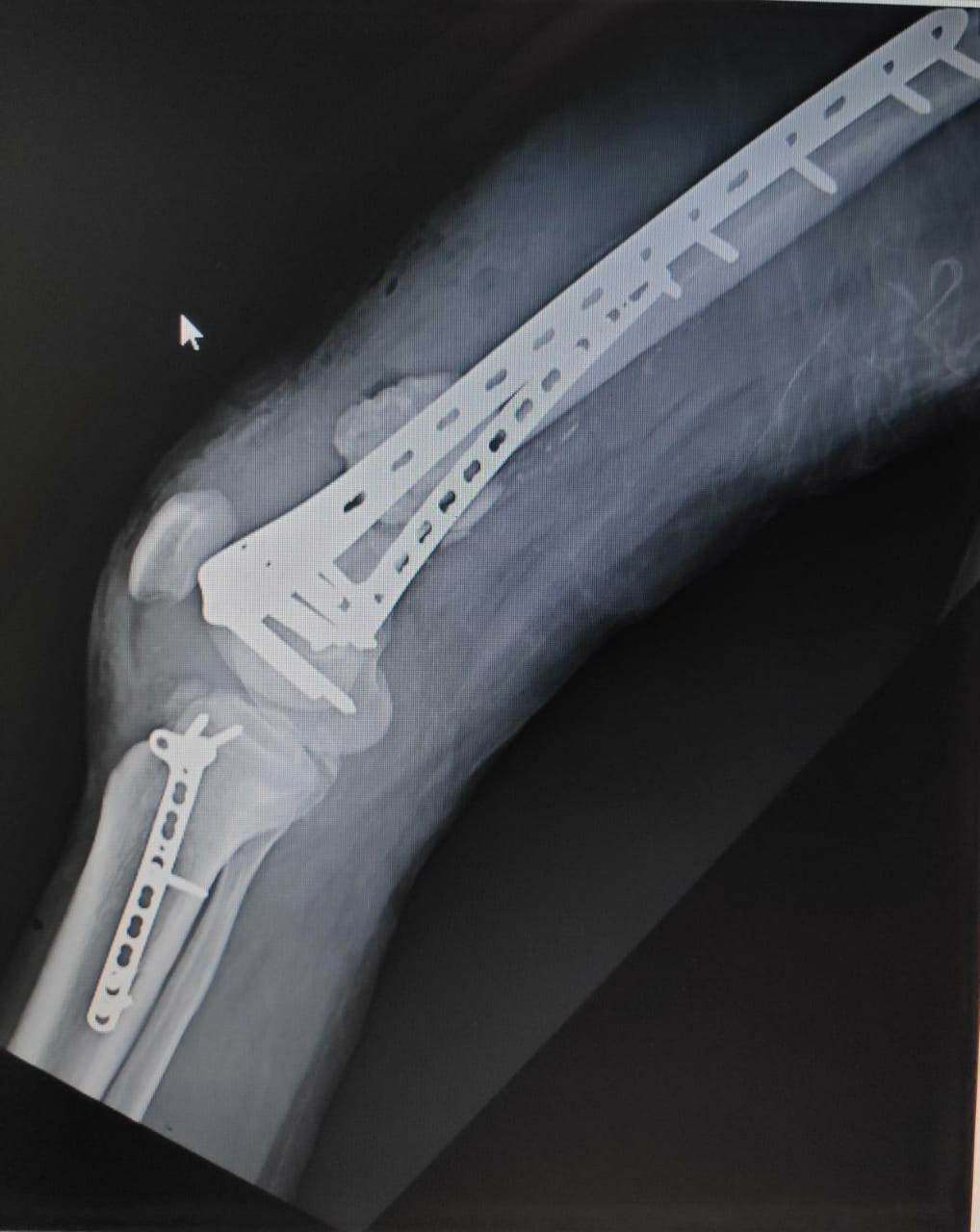
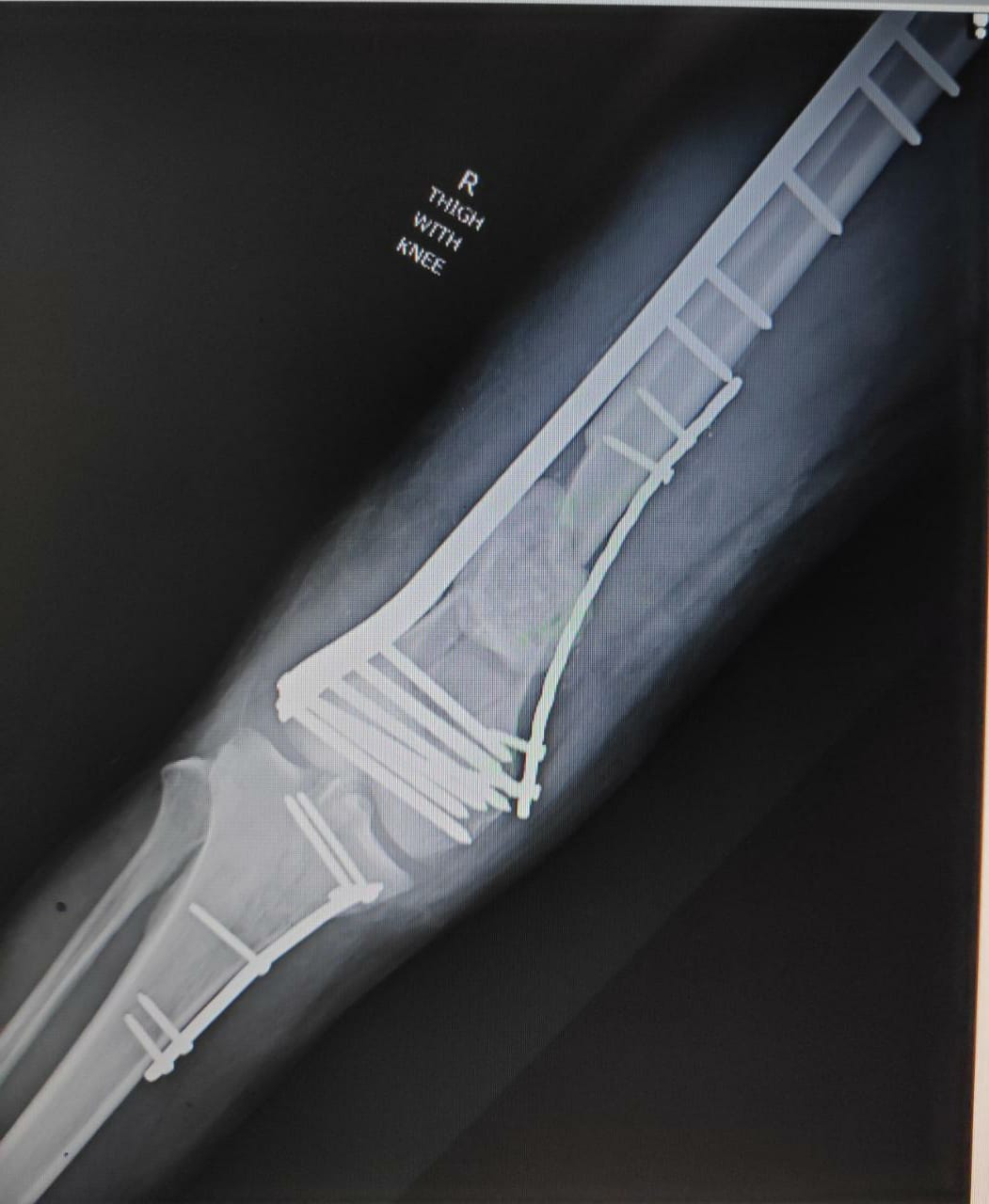
Once serial irrigation and debridement had resulted in a sterile wound, the defect was prepared for the first step of the Masquelet technique. The main aim is to recreate the articular surface of the distal femur. Through direct visualization of the distal femur, a dual plate with screw fixation was done to stabilize the articular surface. Next, an antibiotic-impregnated spacer (preferred to use 3 g vancomycin + 3g of cefazoline per 40 g of cement prepared) was placed in the segmental femur defect and thorough wash was given and an incision was closed in layers with a drain in situ. Once postoperative antibiotics were complete and pain controlled, the patient was discharged from the hospital with non-weight bearing precautions and removal of the drain on the second postoperative day with the advice of knee ROM from postoperative day II.

Stage 2
Six weeks later, the patient was returned with no wound site infection was noticed, X-rays taken and with required investigations, the patient was shifted to operation theatre the pseudomembrane was cleanly incised anteriorly, and the antibiotic spacer was carefully extracted using an osteotome and the fragments were removed. Ipsilateral tri cortico-cancellous strut bone graft from the iliac crest and non-vascularized fibular strut grafts were placed in the defect after freshening the ends, tension-less closure of membrane was done. Tissue sampling sent for microbiological analysis was found negative for any organisms.


Postoperative course
Active & Passive ROM exercises begun from postoperative day 2 with the supervision of a physiotherapist. Active assisted exercises started from the 7th week. The patent was kept on non-weight bearing until the 12th postoperative week, after which partial weight-bearing was started from 3rd month to 6th month, and then to full weight-bearing as tolerated by the patient from the fourth month to 6th month. By the end of the 8th month, the range of motion increased to almost full extension and 100 to 110 degrees of active flexion.

Results
The success of this bony reconstitution allowed the patient to return to daily activities, including full weight-bearing without the need for further assistive devices. Negligible limb length discrepancy was found. All cases showed good alignment in the 6th week. By the end of the 6th-week postoperatively showed early signs of union with evidence of callus formation. The complete union was evident by the end of the 6th-month post bone grafting in 6 patients with no evidence of loosening or infection. Rest all patients had union by the end of the 8th month. All patients retained full weight-bearing by the end of the 8th month.


|
S. No |
Immediate post op |
6 weeks before stage 2 |
3 months post BG |
6 months post BG |
|
Pt no 1 |
0-90 |
0-90 |
0-100 |
0-110 |
|
Pt no 2 |
20-100 |
10-100 |
5-100 |
5-110 |
|
Pt no 3 |
10-80 |
10-90 |
0-100 |
0-110 |
|
Pt no 4 |
10-90 |
5-90 |
5-90 |
5-100 |
|
Pt no 5 |
20-90 |
10-100 |
5-100 |
0-110 |
|
Pt no 6 |
10-100 |
5-100 |
0-105 |
0-110 |
|
Union signs |
6th-week post bg |
3rd months post bg |
6th months post bg |
8th-month post bg |
|
1 |
- |
+ |
+++ |
+++ |
|
2 |
- |
+ |
+++ |
+++ |
|
3 |
- |
+ |
+++ |
+++ |
|
4 |
- |
- |
+ |
++ |
|
5 |
- |
+ |
+++ |
+++ |
|
6 |
- |
+ |
+++ |
+++ |
Discussion
Alain-Charles Masquelet has developed a two-stage technique showing results of 35 patients with diaphyseal defects between 4 to 25cm.[13] Before this Ilizarov and vascularized bone technique was the well-known procedures for large diaphyseal bone defects, this has a longer time to heal and with malposition of the vascularized graft causing deformities.[14] In the Masquelet technique, the reconstruction time is independent of the defect size.[15] Tan et al. described the histology of the pseudomembrane which contains a high amount of mesenchymal stem cells than periosteum in a cohort study of 7-patient, & conclude that because of the high concentration of stem cells which led to the successful reconstruction of large bone defects in this method.[16] Both Masquelet and composite fixation has been successful in reconstructing segmental bone loss independently.[17] We compared our study with previous studies done by Manohar G, Shibu Andrews Study (2012).[18], [19] In his study it showed that fixation with the different plates and its outcome his average flexion and union rate and union time were all more compared to our study with dual plating technique. In another study Siliski[20] the knee rom which was achieved after union was 107° and our study with dual plating clearly showed better results even with gap management. In another study by Rushi Solanki[19] their average rom was 95.8° which is shows our dual plating is far better than their post operatively. Another study by Cheng[21] showed that knee Range of motion can be achieved by orthogonal dual plating fixation. Another study by Zhibiao Bai[22] showed double plating has better outcome and these reports are equal to our findings in our procedures. Another study by Stella[23] showed that dual plating technique allowed the positing of good autograft with good quality which ultimately showed great results. Mohamed A Imam[24] showed that this technique is best for anatomical reduction and stable fixation and it avoids major complications to the patient. Micheal[25] showed union occurred in 12 months which is late than our study which showed far well union than his study. Study by Matthew Gregory Bologna[26] showed that union rate is good in the dual plating than the single plating. Our cases presented unique challenges for surgical planning of reconstruction for limb salvage, the inherent strengths of these two techniques to achieve a successful result. The construct stability which was provided by composite fixation is stable and superior, and our combination of autograft bone succeed within the boundaries of the pseudomembranous capsule which was created by from Masquelet technique. Combining both techniques of reconstruction in an innovative way can be used to achieve even greater successes in future cases with segmental bone loss. Both Masquelet technique and bone grafting are important means in overcoming bone deficits in a trauma patient. In our study, all the patients who underwent the surgery were doing weight-bearing and returned to the regular daily activities after 8 months. None of the cases developed infection nor limb length describes. There were able to perform the rom of the knee from 0 to 110 degrees by the end of the 8th month in 4 cases. Two case showed the late formation of callus and union compared to the others. In this case, they developed flexion deformity of 5 degrees by the end of the 8th month.
Source of Funding
None.
Conflict of Interests
The authors declare that there is no conflict of interests regarding the publication of this paper.
References
- MA Imam, A Torieh, A Matthana. Double plating of intra-articular multifragmentary C3-type distal femoral fractures through the anterior approach. Imam MA, Torieh A, Matthana A:. 121–130, s.l. : springer open article European. J Orthop Surg Traumatol 2017. [Google Scholar] [Crossref]
- EL Steinberg, J Elis, Y Steinberg, M Salai, T Ben-Tov. A double-plating approach to distal femur fracture: A clinical study. Injury 2017. [Google Scholar]
- RM Meneghini, BJ Keyes, KK Reddy, DC Maar. Modern retrograde intramedullary nails versus periarticular locked plates for supracondylar femur fractures after total knee arthroplasty. J Arthroplasty 2014. [Google Scholar] [Crossref]
- PN Streubel, WM Ricci, A Wong, MJ Gardner. Mortality After Distal Femur Fractures in Elderly Patients. Clin Orthop Relat Res 2011. [Google Scholar] [Crossref]
- BL Davison. Varus collapse of comminuted distal femur fractures after open reduction and internal fixation with a lateral condylar buttress plate. Am J Orthop (Belle Mead NJ) 2003. [Google Scholar]
- R Frigg, A Appenzeller, R Christensen, A Frenk, S Gilbert. The development of the distal femur Less Invasive Stabilization System (LISS). Injury 2001. [Google Scholar] [Crossref]
- . Distal femoral fracture fixation utilizing the Less Invasive Stabilization System (L.I.S.S.): the technique and early results. Injury 2001. [Google Scholar] [Crossref]
- JT Watson, M Anders, BR Moed. Management strategies for bone loss in tibial shaft fractures. Clin Orthop Relat Res 1995. [Google Scholar]
- H Weinberg, VG Roth, GC Robin, Y Floman. Early fibular bypass procedures (tibiofibular synostosis) for massive bone loss in war injuries. J Trauma 1979. [Google Scholar]
- . Muscle reconstruction in reconstructive surgery: soft tissue repair and long bone reconstruction. Langenbecks Arch Surg 2003. [Google Scholar] [Crossref]
- AC Masquelet, T Begue. The concept of induced membrane for reconstruction of long bone defects. Orthop Clin N Am 2010. [Google Scholar]
- EA Nicoll. The treatment of gaps in long bones by cancellous insert grafts. J Bone Joint Surg Br 1956. [Google Scholar]
- AC Masquelet, F Fitoussi, T Begue, GP Muller. Reconstruction of the long bones by the induced membrane and spongy autograft. Ann Chir Plast Esthet 2000. [Google Scholar]
- PV Giannoudis, O Faour, T Goff, N Kanakaris, R Dimitriou. Masquelet technique for the treatment of bone defects:tips-tricks and future directions injuries. Injury 2011. [Google Scholar]
- G Owen, T Dyer. Treating Injuries from the War Zone. 2012. [Google Scholar]
- RJ Cuthbert, EA Jones, P Giannoudis, D Mcgonagle, P Giannoudis. The Masquelet technique induces the formation of a mesenchymal stem cell rich periosteum like membrane. 2012. [Google Scholar] [Crossref]
- P Pelissier, A C Masquelet, R Bareille, S Pelissier, J Amedee. Induced membranes secrete growth factors including vascular and osteoinductive factors and could stimulate bone regeneration. J Orthop Res 2006. [Google Scholar]
- SS Siddiqui, S Acharya, J Joshi, R Patel, V Dindod. Functional outcome of orif of distal femur fracture with intra-articular extension. J Evol Med Dent Sci 2014. [Google Scholar]
- R Solanki, A Tolani, S Asati, H Kansara, V Pathria. AO type C distal femur fracture: results of operative management in 52 patients. Int J Orthop Sci 2018. [Google Scholar]
- JM Siliski, M Mahring, HP Hofer. Supracondylar-intercondylar fractures of the femur. Treatment by internal fixation. J Bone Joint Surg Am 1989. [Google Scholar]
- T Cheng, R Xia, X Yan, C Luo. Double-plating fixation of comminuted femoral shaft fractures with concomitant thoracic trauma. Tao Cheng 1, Ronggang Xia 1, Xiaoyu Yan 1, Congfeng Luo 1. 1, Department of Orthopaedic Surgery, Shanghai Sixth People's Hospital affiliated to Shanghai Jiaotong University, People's Republic of China. J Int Med Res 2018. [Google Scholar] [Crossref]
- Z Bai, S Gao, Z Hu, A Liang. Comparison of Clinical Efficacy of Lateral and Lateral and Medial Double-plating Fixation of Distal Femoral Fractures. Sci Rep 2018. [Google Scholar] [Crossref]
- M Stella, E Santolini, A Autuori, L Felli, F Santolini. Masquelet technique to treat a septic nonunion after nailing of a femoral open fracture. Injury 2018. [Google Scholar]
- MA Imam, A Torieh, A Matthana. Double plating of intra-articular multifragmentary C3-type distal femoral fractures through the anterior approach. Eur J Orthop Surg Traumatol 2018. [Google Scholar]
- MA Holzman, BD Hanus, JW Munz, DP O’Connor, MR Brinker. Addition of a Medial Locking Plate to an In Situ Lateral Locking Plate Results in Healing of Distal Femoral Nonunions. Clin Orthop Relat Res 2016. [Google Scholar]
- MG Bologna, MG Claudio, KJ Shields, T Salopek, ER Westrick. Dual plate fixation results in improved union rates in comminuted distal femur fractures compared to single plate fixation. J Orthop 2019. [Google Scholar] [Crossref]