Introduction
Distal enad radius fractures are most commonly found entity in our orthopaedic practice. In adults around 2.4% of total fractures happens contain radius bone fracture and distal end radius fracture constitute 24% of all radius bone fracture.1,2,3 Mode of injury were very different still fall from heights and road traffic accidents being most common mod e of injury. Management of this type of fractures are vary depending upon age of patients, closed or open injury, work profile, life style and surgeon preferences still they all are covered under conservative and operative treatment.4 In conservative treatment like bracing and plaster cast and serial follow ups and mobilization after 1 month. In operative treatment many options are available including k-wire, k-wire fixator, volar or dorsal plate. Tha main objective is to give as much as possible stable and functional joint. Wrist stiffness and weakness in pronation is most common difficulty that we come across. Operative management including volar plating give very much stable fixation of fractures post operatively and we can mobilize wrist as early as possible post operatively.5 Still soft tissue cutting and long mean operative time major drawbacks of this type of surgery that leads to non union of fractures and post operative infections some times. For better fracture site exposure we have to separate pronator quadratus muscle from the radius side so there is different opinions of surgeons regarding repair of pronator quadratus muscle. If we try to repair pronator quadratus after plate fixation than it covers implant and thus it reduce friction injury to volar tendons.
We have evaluated clinical, physiological, radiologic outcome in our retrospective study regarding distal radial fractures operated with volar plating in which some patients had PQ repair done and some patients without PQ repair.
Materials and Methods
This randomized retrospective clinical study was conducted on 30 patients with distal radial fracture treated with volar plating by trained single surgeon in the Department of Orthopaedics, B.J. medical college, Civil hospital, Ahmedabad between January 2017 and January 2019. These cases were selected for study randomly as came during opd practice and in emergency. Fracture patterns were classified on basis of AO/OTA classification of distal radial fractures.
Exclusion criteria
Patient’s Age < 18 yr. - > 60 yr.
Open fracture
Osteoporotic patients
Pathological fractures
Associated other fracture
30 randomised patients having distal end radial fractures presenting within 10 days of clinical injury and operated with the volar plating between January 2017 to January 2019 were included in this retrospective study. Mean age of this patients included in study was 44 years (range 18–60 years). The mode of injury included road traffic accident in 15(50%), fall from height in 14(47%), sports injury in 1(3%).
For fracture classification AO classification for distal end raidus was used. All patients were operated in supine position with using tour niquettied at arm level and pressure set to 250mm. Open reduction and internal fixation was used for all volar distal end radius fracture.6,7 All operations were performed by orthopaedic surgeon of civil hospital ahmedabad. A standard volar approach was used for plating. After 1st dressing in 24 hr. post operatively when pain was decreased assisted passive mobilization of wrist was started and physiotherapy of wrist and elbow was explained. Assisted excercises and active physiotherapy was started after 1 month of post operative period. All patients were followed of after 15 days for suture removal and than again after 15 days for x-ray evaluation. Than after monthly followed up were taken to see union of fracture on xray showing callus formation. Each and every patients were evaluated clinically for range of motion at wrist joint and followed up for visual analogue scale and patients satisfaction. Daily activity and light weight lifting was allowed after one month of follow up and heavy weight lifting was allowed after 3 month of post operative period.
Surgical Technique
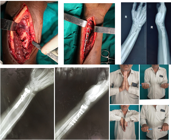
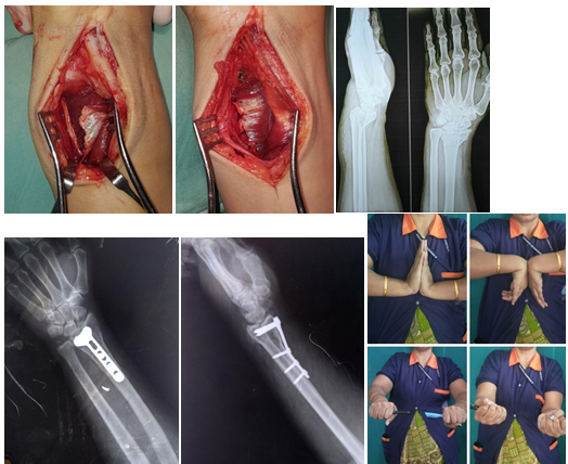
All patients were shifted to operation table and after patient got induced supine positi on was given with injured upper limb was painted and drapped with tourniquet held on upper arm. Volar approach (modified henry approach) was used by palpating flexor carpi radialis tendon and skin incision was done over anterier aspect of forearm. To reach fracture dorsal site pronator quadratus was detached from border of radius through L shape manner. After fracture reduction with traction and manipulation under IITV guide, volar plating was done and plate was fixed with bone with screw and in some patients pronator quadratus muscle was repaired with synthetic absorbable suture and in some patients pronator quadratus was kept as we had cut. On 1st post operative day passive exercise were started under physiotherapy assistance. And after suture removal active and passive all movements and mobilization was started and light weight bearing was allowed after 6 weeks.
Observations and Results
Total 30 patients having distal end radial fracture managed with volar plating were selected with mean age of patients was 44yrs (18-60yrs) and out of them 22 were male while 8 were female. In all of them, mode of injury was fall down from heights and RTA. Mean operation time was 04 days from admission date. Average operating time was 60 ± 10.5 min (30–95 min) for volar plating. Hospital stay varied from 5 days to 14 days, mean being 9 days. Average time of callus formation and seen on xray was found to be 21 weeks.
The mean operative surgical time was approximately 60 ± 10.5 min (30 – 95 min) for radius volar plating. All patients were regularly followed up in hospital and mean followed up time was 13 months (range from 06 to 20 months). Out of 30 patients having distal end radius fracture, 28 patient’s (94%) fractures were united with mean time around 21 weeks (range 16-29 weeks ). One patient (3%) have non-union at fracture site which require revision surgery with bone grafting. One patient (3%) having post-operative infection which is covered with higher antibiotics and regular dressing, no need ed to revise surgery. No patients found any plate impingement hence no implant removal needed. Almost 95% patients have excellent to good palmar flexion prest at wrist joint in long term follow up. Mostly patients have range from 65-85 degrees and average was taken as 75 degree.
All the patients have dorsi flexion ranging from 65 to 92 degrees and average was taken as 82 degree for dorsi flexion. Around 96% patients have excellent to very good supination range at wrist joint ranging from 76-86 degrees and average was taken as 80 degree. All patients have excellent to very good pronation range at wrist joint ranging from 80-90 degrees and average was taken as 85 degree.
In our study, 97% patients have excellent radial deviation range at wrist in long term follow up. Most of patients have around 11-19 degrees radial deviation and average was taken as 11 degree. Most of patients have excellent ulnar deviation range at wrist in long term follow up. Most of patients have around 20-30 degrees ulnar deviation and average was taken as 25 degree.
X -rays was taken to evaluate radial inclination, radial shortning and palmer tilt. in our study athe average radial inclination is 20 degrees, average radial length is 11mm, average pal mertilt is 7 degrees. Around 93.33 % of patients had excellent to good outcome according to gartland and werley score.
Table 1
Criteria for mayo wrist performance score
Table 2
| Interpreting the Mayo wrist Performance Score | |
| 90 to 100 | Excellent |
| 80 to 89 | Good |
| 65 to 79 | Fair |
| <65 | Poor |
Intrpretation of mayo wrist performance score
Results
Table 5
| G&W Score | Number of patients | Percentage |
| Excellent 0-2 | 24 | 80% |
| Good 3-8 | 4 | 13.33% |
| Fair 9-20 | 2 | 6.66% |
| Poor >20 | 0 | 0% |
| Total | 30 | 100% |
Scoring system (Gartland & Werley (G & W) Score)
Table 6
| Palmer flexion | Dorsi flexion | pronation | Supination | Radial deviation | Ulnar deviation | |
| Average | 75 | 82 | 85 | 80 | 11 | 25 |
Clinical outcome
Discussion
Distal end radius fracture are commo n fracture of upper extrimites, various modalities are available like k wire, k-wire with external fixator, volar plating, now more commonly volar plating is the perfomed surgery for the same.8 Patients managed with external fixation have more complication like distal radius ulna joint stiffness, decreased supinaton pronation strength, lack of stability than patients managed with open reduction and internal fixation.8,9 But to perform volar plating surgery, the detachment of pronator quadratus muscle is needed to expose fracture site, so after fracture fixation with volar plate deliema lie in betweeen repair of pronator quadratus should be done or not, and repair of pronator quadratus muscle will cover the implants hence friction injury to flexor tendons can be prevented and full pronation movements can be achieved.10 Same in every surgery there are some risk or disadvantages like malunion, loss of fracture reduction, post operative infection, stiffness of joint and sometime skin problems. This study is conducted to assessed the out come of both the patients groups operated with distal end radius plating with PQ repair done and without PQ repair. Some surgeon are in favour of repair of PQ muscle that leads to give well pronation strengths in range of motion of wrist joint in post-operative period. Still multiple parameters were taken into consideration to evaluate and compare results.
In our study we found that there is Little difference in pronation strength post operatively still after 10 week of surgery there is no difference found in pronation strength and active range of motion in wrist joint. All the patients can carry out there routine work with out and difficulties. Still we suggest to take longer follow-up of all patients. Furthermore, we have also compared the strength and movements from operated limb to other non operated limb of same person and we found that initially up to 9 month there is some less isometric pronation strength as compared to non operated limb but after 12 month post operatively there is no significant difference in movements and strength anymore. So practically dissection of PQ have no or minimal impact on forearm pronation function in a longer way. Trosti and Ilyas had conducted a prospective evaluation study of pronator quadratus repair versus no repair in patients operated with volar distal radius plate fixation with follow-up of 12 months post opertively. They have even found no significantly different results between these two groups according to Range of motion at the wrist joint, DASH scores, VAS scores, and grip strength.11 Knirk JL also conducted outcome and prognosis study on over 112 patients operated with volar radius plating with pronator quadratus repair and non repair and did follow up for 1 year post operatively and he didn’t found any signific ant differences in pronation at wrist joint and pain at wrist joint according to his study there is no further advantage in repairing the pronator quadratus muscle post operatively after volar distal radius plating.12 In prospective study of Chirpaz-Cerbat JM shows that pronator quadratus sectioning required by the anterior approach entails a risk of pronation strength loss and of distal radioulnar joint destabilization still long term follow up shows no significant difference whether pronator quadratus repaired or not.13
However the repair of pronator quadrarus muscle can be challenging as sometime there is some traumatic disruption is there and due to poor quality soft tissue and in old patients. So in our set up we try to repair PQ if it is possible but still there is no longer clinical benefits in strength or in movements found. We found only benefits like reduced pain in early post-operative period. And it covers implant so decreased friction to flexor tendons so tendon injury can be prevented.
Conclusion
After pronator quadratus repair pain reduced in early postoperative period but pronation strength in early rehabilitation period could not confirmed, And both group of patients give satisfactory and almost similar results with full range of movements and early mobilization and almost same pronation strength in longer follow up. We use and recommened to repair pronator quadratus muscle to cover plate and protect flexor tendons from friction injury due to plate and screw heads. In our stuy we found repair of pronator quadratus muscle will give benefits for early post operative period but longer follow up shows no clinical difference in supination and pronation strength.


