Introduction
Osteoarthrosis of knee is the most common cause of disability in the older.1 Pain and limitation in the mobility of knee are the major causes of disability.1 Total knee arthroplasty, which aims to relieve pain, improve joint function and mobility, is the main surgical treatment in this patient population.1 The First choice of treatment in the young patients with medial compartment osteoarthrosis is high tibial osteotomy even though this is associated with some disadvantages like non-union.2,3,4,5 Proximal fibular osteotomy is the new alternative in the young patients which is safe, simple and affordable1. It relieves pain immediately and delays or avoids the need for total knee replacement.1 The procedure is to remove 1.5 to 2.0 cms piece of fibula Seven to 10 cms below the head of fibula to relieve medial compartment pressure, and consequently realign the knee.
It was proposed that the medial part of the knee has only single cortex support in an otherwise fully cancelous bone which is insufficient over a period of decades with mild collapse with ageing while the lateral part of the knee however is s upported by three cortices (one by tibia and two from fibula) making it rigid and less collapsible. This leads to increasing varus with ageing and causes medial compartment osteoarthrosis of knee.6 It was also believed that load is distributed medial to centre of knee along mechancal axis.7
Materials and Methods
Instituitional Ethics Commitee (IEC) approval obtained for this study (No.ECR/1160/Inst/ap/2018). All patients were given informed consent and agreed to participate in the study.
From July 2016 to Jul y 2017, 60 consecutive patients who underwent PFO at our hospital were followed prospectively (n = 37; age range, 48–72 years; 40 female, 20 male) with follow- up at 3,6,9 weeks, 3 months, 6months, 9 months, 12 months, and 24 months post- operative. Outcome measured by femorotibial angle using wang et al method8 and pain using visual analog scale.9 The diagnosis of osteoarthrosis was made by the clinician according to the American College of Rheumatology criteria.10
Inclusion Criteria
Isolated medial compartment arthritis of the knee with a varus deformity not exceeding 12 degrees.
Normal lateral compartment
Near normal Patellofemoral joint and
At least 2 mm joint space in the medial compartment, on weight bearing X-rays.
(Patients suitable for uni -compartmental arthroplasty or HTO)
Details of procedure
With patient under spinal anesthesia and in supine position a longituidinal incision of about Five cms made over skin of the proximal fibula. Periosteum over the fibula is exposed between the peroneal and soleus muscles. Proximal fibular osteotomy was made about Seven to Ten cms from head of the fibula by removing about One to Two cms length of fibula.
With in few hours after the surgery patient is allowed full weight bearing and mobilisation of the knee. Weight bearing radiographs of the knee taken post operatively and compared with pre operative radiographs. Medial and lateral joint spaces analyzed and amount of correction achieved is determined. Knee pain was assessed using VAS (visual analogue scale).
A mbulation activities of knee were recorded using the knee and function subscores of the American Knee Society score11 preoperatively and a mean of months postoperatively. Statistical analysis performed with mean +SD for continuous variables.
Results
Out of 60 patients who underwent PFO medial pain relief was observed in 54 patients after PFO with in one or two days post operatively remaining 6 had no change in pain. The mean duration of follow-up was 2 years (range,12 months). The average duration for unilateral PFO was 32.23+/- 9.13 minutes. Postoperative complications like paresthesias on dor sum of foot were observed in 6 patient s, which recovered by 6 weeks to 12 weeks and no other complications including wound infection, delayed healing or nerve damage.
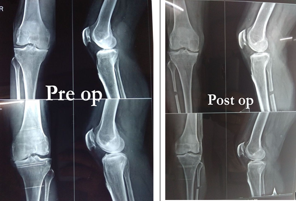
Figure 6
2 more patients xrays showing improvement in alignment and increase in medial joint space post operativel

The mean knee and function subscores were (American Knee Society score) .
Table 2
| Pre operative | 44.41 +/-8.90 and 41.24 +/-13.48 |
| Post operative | 69.02+/- 11.12 and 67.63 +/-13.65 |
Radiographs of the weight-bearing lower extremity showed average increase in the medial knee joint space postoperatively compared with preoperatively. The ratio of the knee joint space in cms (medial/lateral compartment).
Discussion
Proximal fibular osteotomy is a safe, simple and affordable day care surgery that doesn’t require insertion of implants. PFO is thus one of the best alternative for osteoarthritis of medial compartment of knee especially for patients with medical comorbidities who cannot undergo arthroplasty.
In our study 54 cases were successful (out of which 6 patients got nerve complaints (tingling and numbnes) who were treated with Pregabalin 150 mg and vitam in b12 for 6 weeks to 12 weeks for these nerve complaints to settle) and 6 patients had no significant relief of pain in the followup period.
As the procedure leaves the knee virgin and is performed away from the knee joint, it scores over H.T.O in not violating the joint for a subsequent TKA if needed.
Conclusion
Thus we conclude that proximal fibular osteotomy is a safe, simple and afford able day care surgery emerging as an alternative for high tibial osteotomy for patients with arthritis of medial compartment. Also further studies with higher level of evidence needed for confirmation of findings.


