Introduction
Pigmented Villonodular Cynovitis (PVNS) is a rare disease affecting the joints which are lined by synovium. It was described by Jaffe and colleagues, in 1941 as a benign inflammatory state of the synovium unclear etiology and as a tumor like aggression of synovial tissue involving joints.1 It can be either diffuse or localized either intra articular or extra articular.2 Diffuse PVNS involves the entire joint synovium whereas localized PVNS is a discrete nodular, lobulated mass.3 PVNS mainly occurs in the knee joint, PVNS of the foot and ankle is rare4 (incidence, is approximately 1.8/million.5 Severity of bony involvement in PVNS of the ankle is high, because the pressure erosion easily occurs in the narrow joint space of the ankle joint.6 The diffuse type has high recurrence rate as high as 50%, whereas the rate for the localized type is considered low.[2,5,7]2,5,7 To prevent, complete excision and careful tissue handling is necessary. We operated a case of ankle(talo calcaneal) PVNS with bone erosion of adjoining articular cartilage of talus and calcaneus which was treated with radical excision. The authors have obtained the patient’s written informed consent for print and electronic publication of the case report.
Case Report
38 year old male patient was referred to us with complains of pain, gradually increasing swelling and ulcerating wound on lateral aspect of right ankle since 10yrs with history of excision done in past. On examination swelling was present on lateral aspect of right ankle range of motion and stability of joint appeared to be normal. USG of local area was done which showed approximately 7*3.5*8 cm sized well defined heterogeneous isoechoic lesion. A true cut biopsy was taken which showed loosely cellular fibro vascular tissue with diffusely infiltrating lymphocytes and many brown pigmented laden cells and vascular proliferation. Diagnosis of pigmented villonodular synovitis was done. Surgical resection of mass was planned with diagnostic arthroscopy t o see for extent of the tumor with standard J-Shaped inci sion. The challenges which we faced in surgery was wound closure, risk of infection, Paraesthesia involving the distribution of superficial peroneal nerve.
Figure 2
Plain radiograph of ankle showed shadow of the swelling and confirmed that the swelling was not bony

Figure 3
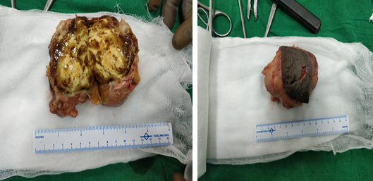
Histopathological sample revealed grossly a greyish brown with few white areas (? Pigmentation) soft to firm in consistency and encapsulated tissue of size 3.5*2.8*1.2 cm

Discussion
PVNS is mainly affects of synovial joints. PVNS rarely occurs around ankle joint<10% as compared to other joint and no standard treatment has been defined. Arthoscopic or open arthotomy can be performed. Treatment of choice is synovectomy for diffuse PVNS, because of high recurrence trails for use of adjuvant focused external radiotherapy with intra articular injection with radioactive colloids.
Total synovectomy and complete excision is necessary to prevent joint erosion and degenerative changes. Because of anatomic characteristic of ankle joint it is technically difficult to exfoliate massive tissue without disturbing integrated ligaments. In our case the patient had massive mass extending to talocalcaneal joints with involvement of articular cartilage. Thus to summarize the localised PVNS involving the ankle joint needs complete mass excission


