Introduction
An osteochondroma is a benign tumour that consists of a bony outgrowth connected to the cortex and medullary cavity of the parent bone and covered by a cap of hyaline cartilage, It occurs most frequently in the metaphysis of long bones such as the proximal humerus, proximal tibia, distal femur, and in the pelvis.1,2,3,4 It is usually asymptomatic and diagnosed accidentally during routine radiographic examination,3,5 However it may become painful when it impinges on the surrounding structures or joints.
Osteochondroma is the most common benign tumor of the bone,4,6 accounting for about 20-50% of all benign bone tumors and 10-15% of all bone tumors.6,7 However, it is uncommon around the foot and ankle, and rare in the talus, the Scottish bone tumor registry identified only 23 bone tumours arising from the talus over 56 years follow up starting from 1954 to 2010.8 Of these 23 cases, 3 were malignant, 20 were benign and only 2 cases were osteochondoma. Therefore, The presentation of osteochondroma in the talus is very rare and only few cases had been reported,4,5,9,10,11,12 we report a 9-year-old male with a large painful osteochondroma of the talar neck, part of it was broken and separated as an intra articular loose body, the other part was connected to the talar neck by a stalk and it was successfully treated by surgical excision.
Case Report
A 9-year-old male primary school student presented with a 1 year history of swelling over his right ankle which was gradually increasing in size. The ankle had started to be painful 6 months earlier, the pain was activity related during walking and climbing up stairs, it interfered with running and sporting activities and he had difficulties with fitting shoes. The mass was enlarging insidiously and there was a history of mild trauma from which the patient recovered within 2 weeks of rehabilitation but the patient continued to experience activity related pain.
Physical examination revealed a hard mass approximately 5 × 4 cm anterior to the ankle joint just lateral the medial malleolus, the mass was attached to the underlying bone, but not fixed to the overlying skin, with globular surface, there was limited inversion and ankle dorsiflexion was limited to 10 degrees with no ankle instability and no limb length discrepancy.
The posterior tibial and dorsalis pedis pulses were palpable and of a good volume with no neurological deficit or muscular weakness around the ankle and no regional lymphadenopathy, there were no other palpable masses in the other limbs.
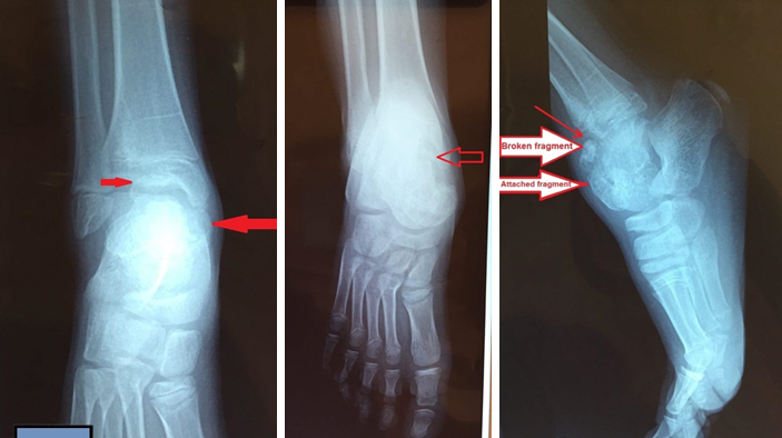
Anteroposterior and lateral ankle radiographs revealed a large pedunculated bony mass originating from the anteromedial aspect of the talar neck and a smaller bony mass which appeared to be free or broken off the bigger mass (Figure 1). There was a small defect on the anterior edge of distal tibia epiphysis which could be due to pressure and repetitive impingement by the mass.
A provisional diagnosis of osteochondroma or synovial chondromatosis had been made and surgical excision decided as the mass was painful and was associated with anterior ankle impingement.
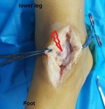
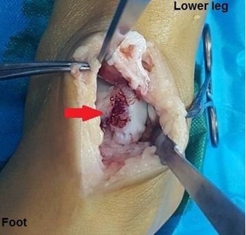
The patient was placed supine, and the operation was carried out through anteromedial approach. The tendons and neurovascular structures were retracted, and a small 2.5 ×1.5 cm loose bony mass was taken out which was covered by hyaline cartilage, the larger 3.5×2.5 cm bony mass was found to be attached to the talar neck by a pedicle, it was separated by osteotomy and the entire mass was excised (Figure 2,Figure 3,Figure 4). The wound was closed with interrupted non-absorbable sutures.
Histopathological examination of the tumour confirmed the diagnosis of osteochondroma with trabecular bone, adipose tissue of the marrow and a surrounding hyaline cartilage cap. There was no evidence of malignancy.
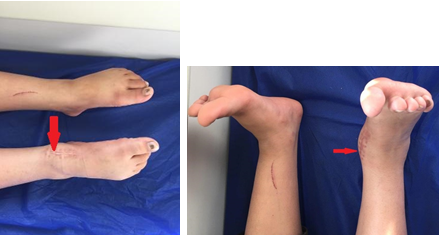
The patient had an uneventful postoperative recovery, at 8 weeks postoperatively he was free of pain with healed wound (Figure 5) and at 10 months he had regained full range of motion, participated in sporting activities with no signs of recurrence. Post operative radiograph showed complete resection of the tumour (Figure 6).
Figure 1
Anteroposterior radiograph (A& B) of the ankle demonstrating the mass . lateral radiograph (c) showing the broken and attached fragments of the mass ,and the indentation on the distal tibial epiphysis caused by the anterior impingement with the mass.

Figure 2
Intraoperative photograph, antromedial view showing the attached part of the osteochondroma arising from the talar neck.

Figure 3
Intraopeative photograph, antromedial view of the right ankle after excision of the osteochondroma showing the talar neck.

Figure 5
Photograph of the ankle 8 weeks postoperative, showing healed wound and near full range of motion of the ankle.

Discussion
Osteochondromas are the most frequently seen benign bone tumors,4,6 However, it is quite unusual for them to originate from the foot and ankle region, especially from the talus.4,5,8,9 Ozdemir et al13 in a retrospective study found out that only 196 tumours out of 1786 bone and soft tissue tumor were located in foot and ank le. Osteochondroma of the foot and ankle arises mostly from the phalanges and metatarsal bones, and it is very uncommon to present in the talus.4,9,10,11,12 Osteochondromas of the talus are usually located on the dorsal surface,9,10,11 they are usually asymptomatic and diagnosed incidentally. However, it may give rise to symptoms such as pain, limitation of motion, a mass effect, and ankle swelling. Pain occurs when it reaches a large size or causes impingement on the surrounding tendons and joints or when it gets fractured.14 A fracture in an osteochondroma which commonly occur through the stalk could be the result of a blow, or indirect muscle or tendon injury as described by Carpintero et al.14 Our patient gave a history of minor trauma to the ankle which could explain the separated small fragment of the osteochondroma that was shown both by preoperative radiographs and intraoperatively, unlike the previous reports14 the fracture was not through the stalk.
While accidentally discovered osteochondromas in asymptomatic individuals are managed with observation, Resection is indicated for patients with a painful mass secondary to irritation of the surrounding soft tissue, for a lesion in a location that is prone to repetitive trauma, for a lesion associated with cosmetic deformity or potential damage to nearby joints or neurovascular structures, and for a lesion that has features of malignant transformation.15,16,17 The patient should be informed about the rare possibility of malignant transformation (<1%) especially when there is new onset of symptoms or rapid enlargement.18 Plastic deformation of tibia, mechanical blocking of joint motion are other justifications for surgical resection, therefore, most of the authors choose a surgical resection for osteochondromas in this region5
In the present report, we have described a case of talar osteochondroma with all the typical symptoms like pain, swelling, mass effect and limitation of his left ankle functions and daily activities, there was a history of minor trauma to the ankle but it cannot be confirmed whether the fracture in the osteochondoma was due to that trauma or not ,although most cases of osteochondroma could be managed conservatively, surgical resection is required when it becomes symptomatic and interferes with normal ankle function as in our case, resection rendered the patient free of symptoms with resolution of the swelling and gradual return of full range of motion of the ankle and return to sporting activities.
In conclusion osteochondrom should be put on the list of differential diagnosis of a mass around the ankle whether it is painful or not, surgical resection with thorough removal of tumour is the most successful treatment of symptomatic osteochondroma.


