- Visibility 19 Views
- Downloads 4 Downloads
- DOI 10.18231/j.ijos.2020.021
-
CrossMark
- Citation
Functional outcome of modified Blair’s arthrodesis for injuries of Talus
- Author Details:
-
Srinivasa Rao Akula
-
Sandeep Vella
-
Sai Sowmya Bondili *
Introduction
Fracture neck of talus associated with or without sub talar, tibio talar and talo-navicular joint dislocation can be considered as serious injuries around ankle and poses challenging problem for an orthopaedician when these fractures are neglected and resulting in non-union and osteonecrosis of the talus. Talar neck fracture and body dislocation occurs due to forced ankle dorsiflexion combined with axial loading, results in talar neck impacting against the anterior edge of tibia. These fractures sometimes can be treated with manipulation and casting. Detenbeck and Kelly recommended excision of talus and tibio calcaneal arthrodesis by compression as main stay of treatment for fracture dislocation around talus but this technique resulted in widening of hind foot and shortening of foot which lead to difficulties in shoe fitting.[1] To avoid this complication Blair performed a tibiotalar fusion, in this procedure body of talus was removed and sliding cortical tibial bone graft was placed anteriorly between anterior aspect of tibia and head of talus.[2] After that Morris et al. modified the procedure by introducing a trans-calcaneal Steinmann pin through calcaneum in to tibia and fixing tibial sliding graft with a cortical screw proximally to prevent proximal migration of sliding graft.[3] With this procedure the foot maintained its normal appearance, alignment and shortening was minimized and remaining subtalar range of movements were increased.
The present study evaluates the functional outcome of patients treated by modified blair’s arthrodesis using American orthopaedic foot and ankle society score and visual analog scale preoperatively and postoperatively and tibio pedal movement postoperatively.
Materials and Methods
This is a prospective study conducted in katuri medical college and hospital, Guntur, from July 2018 to January 2020. Total nine cases who were treated with modified blair’s arthrodesis for non-union and osteonecrosis were included in this study. The median interval between injury and index surgery was 6 weeks to 20 weeks. Among nine patients, 6 (66.6%) were males and 3(33.3%) were females. Preoperatively roentgenogram was performed to all patients for conformation of fracture and were managed with modified blair’s arthrodesis using sliding graft from anterior tibia. All the patients were followed up for 1 year with serial radiographs and tibio pedal movements were assessed postoperatively American orthopaedic foot and ankle society score and visual analog scale were assessed preoperatively and at postoperative 1 year follow up.
Statistical analysis
Statistical analysis was performed using Statistical package for social sciences 23.0 medical software package. Data were analyzed with t- test. P<0.05 was considered statistically significant.
Surgical procedure
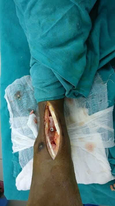
Through anterior approach, interval is developed between extensor hallucis longus & extensor digitorum longus. After exposure of joint, tibial articular surface is denuded, foot placed in 0 degrees of dorsiflexion, 5 degrees valgus and 10 degrees of external rotation and tibial, talar surfaces are placed in contact with each other and sliding bone graft of size 5 cm × 2.5 cm from anterior aspect of tibia is inserted in to talar neck and fix the proximal part of graft with a 4.5mm cortical screw. To increase stability at arthrodesis site and to prevent varus and valgus deformity of foot a trans calcaneal Steinmann pin is passed through calcaneum in to tibia.

Post-operative management
Postoperatively, long leg cast with knee in 30 degrees flexion was applied for 6 weeks. After 6 weeks, Steinmann pin removal was done and short leg walking cast was substituted. Non weight bearing walking up to 12 weeks after surgery and weight bearing is allowed after healing of graft which is confirmed by radiography. Tibio pedal movement was assessed clinically. Tibio pedal movement is defined as arc of movement from maximum dorsiflexion to maximum plantar flexion, and angles subtended by long axis of tibia and foot in lateral projection.
Results
Practically, results were assessed based on degree of tibio pedal movement and ability of patient to do full activities without any symptoms. We have considered the patient have had an EXCELLENT result if degree of tibio pedal movement is of 15-20˚ and is able to do his daily activities without any symptoms and outcome of patient is considered to be GOOD if the patient is having tibio pedal movement of 10-15˚ and have occasional discomfort but no restriction of daily activities. Outcome is considered to be poor if tibio pedal movement is less than 10˚and daily activities of patient are restricted because of severe pain. Final outcome was shown in ([Table 1]).
| Final out come | No of patients | Percentage |
| Excellent | 5 | 55.5% |
| Good | 4 | 44.4% |
| Poor | 0 | 0 |
The position of arthrodesis of ankle was assessed clinically and is fused in 0 degree dorsiflexion in all cases and fusion was assessed radiologically by formation of trabeculations across tibia, talus and sliding graft. All the patients were with satisfied ankle function postoperatively. American orthopaedic foot and ankle society score (AOFAS), visual analog scale scores (VAS) were significantly different 1 year after surgery compared to preoperative period (p<0.05), indicating significant improvement in ankle joint function ([Table 2]).
| Item | *AOFAS | *VAS |
| Preoperative | 51.3±16.19 | 2.89±1.05 |
| Postoperative | 85.44±5.20 | 0.44±0.88 |
| T | 6.13 | 5.5 |
| P-Value | 0.0003 statistically significant | 0.0001 statistically significant |
On measuring limb length no shortening was noted. Normal heel height and shape were maintained in all the cases. Gait was assessed which is physiologically normal. No limp was noticed and patient is able to walk more physiologically.


Discussion
The best suggested treatment of fracture in non-union and Osteonecrosis of talus is blair’s arthrodesis. In 1943 blair described tibiotalar arthrodesis as main stay of treatment for fracture of the talus in non-union and osteonecrosis.[2] This technique involves removal of fragments of talar body and insertion of sliding tibial graft in to neck of talus. In 1971 morris et al. modified this technique by fixing the graft to tibia with a screw proximally and tibia on to calcaneum is fixed with transcalcaneal Steinmann pin.[3] These modifications stabilized the ankle. Linsy et al. and Patterson et al. performed modified blair’s fusion, but none of them tried to salvage talus body.[4] Kitaoka et al. explained about talar body removal for first time.[5] These studies stated that when blair’s arthrodesis is performed with excision of talar body, loads of three to four times body weight occurs in ankle joint during normal walking. With removal of posterior facet, forces acts on subtalar joint and contact characteristics of anterior and middle facet changes substantially leading to degenerative arthritis of sub talar joint. In this study, bone is not excised instead tibial articular surface is denuded which increases not only the intraoperative stability but chances of varus and valgus are reduced. With this technique, cortical bone with added cancellous bone from lower end of tibia produces sound fusion in all cases and also maintains normal height of heel.
Most of the patients had a successful clinical result, almost the normal alignment of the foot with relative to ankle and leg was observed. Radiologically the talus, the graft and tibia got incorporated and sound fusion was achieved in all the cases. Dunn et al. was first to observe the revascularization of the talar body after fracture and it took many years to for revascularization.[6]
| Study | Preop AOFAS | Postop AOFAS | Preop VAS | Postop VAS |
| Wang Shuangli et al. | 45.38±3.21 | 83.13±3.76 | 8.01±0.63 | 2.31±1.05 |
| Shay Tenenbaum et al. | 32.7 ± 8.7 | 72.1±10.1 | 6.9 ±1.5 | 1.7± 2.2 |
| Our study | 51.3±16.19 | 85.44±5.20 | 2.89±1.05 | 0.44±0.88 |
For normal physiological gait 10-20° of tibiopedal movement is necessary.[7] With limitation of arthrodesis at tibiotalar level and preservation of talus, hind foot function remains unaltered by sharing load to anterior and middle facet of subtalar joint. In the present study four patients (44.4%) showed tibio pedal motion of 10˚- 15˚ and five patients (55.5%) showed tibio pedal motion of 15˚-20˚, resulting in good to excellent gait. There was no certainty in position of arthrodesis as varied opinions among different authors regarding best position for arthrodesis was noted. Barr and record recommended 5˚equinus,[8] Watson-jones as 10˚ equinus for arthrodesis.[9] Anthony A. Mascioli preferred neutral dorsiflexion as best position for arthrodesis.[10] In this study the ankles were fused in 0 degree dorsiflexion with which patient is able to walk more physiologically without any difficulty. With this technique, AOFAS was significantly increased and VAS was reduced.
The comparative functional outcomes of pre-Op and post-Op AOFAS and scores of various studies performed over the years shown in ([Table 3]).
Conclusion
We achieved good longterm results with modified Blair’s arthrodesis. Arthrodesis with preservation of talus provided greater intraoperative stability and almost normal looking foot with no limb length discrepancy. This technique has high reliability in pain relief and remained tibio pedal movement helps the patient to walk more physiologically without any difficulty.
Sources of Funding
None.
Conflict of Interest
None.
References
- L C. Detenbeck, P J Kelly. Total Dislocation of the Talus. J Bone Jt Surg 1969. [Google Scholar]
- Harry C. Blair. Comminuted fractures and fracture dislocations of the body of the astragalus: operative treatment. Am J Surg 1943. [Google Scholar]
- H D Morris, W L Hand, A W Dunn. The Modified Blair Fusion for Fractures of the Talus. J Bone Jt Surg 1971. [Google Scholar]
- Brendan M. Patterson, Allan E. Inglis, Bruce H. Moeckel. Anterior Sliding Graft for Tibiotalar Arthrodesis. Foot Ankle Int 1997. [Google Scholar]
- H B Kitaoka, G L Patzer. Arthrodesis for the Treatment of Arthrosis of the Ankle and Osteonecrosis of the Talus*. J Bone Jt Surg 1998. [Google Scholar]
- Allan R. Dunn, Bernard Jacobs, Rolla D. Campbell. Fractures of the talus. J Trauma: Inj, Infecti Crit Care 1966. [Google Scholar]
- T Waugh. . Joe King Visiting Lectureship. Baylor College of Medicine 1977. [Google Scholar]
- J S Barr, E E Record. Arthrodesis of the ankle joint: indications, operative technic and clinical experience. New England Journal of Medicine 1953. [Google Scholar]
- R Watson-Jones, E. S. Livingstone. . Fractures and joint injuries 1962. [Google Scholar]
- F M Azar, S T Canale, J H Beaty. . Campbell's operative orthopaedics e-book. Elsevier Health Sciences 2016. [Google Scholar]
