Introduction
The ankle is a hinj joint and having two articulation points, 1) between distal parts of tibia and fibula and 2) between tibia and talus. The ankle joint is most important joint for weight bearing purpose for which supportive movement occur between sub talur joint(between talus and calcaneum) and at ankle joint. Mostly weight is transferred by tibio talar surface and only 1/6 part of weight is transferred by fibula side. Thus mortise view is very important in case of any fracture management of ankle joint as it shows talur dome so greatly that weight bearing results can be assessed properly. Movement in ankle joint occure mainly at sagittal axis and also in longitudinal and empirical axis.1
Centre of ankle joint is 3-4 mm lateral to centre of inter malleolar axis which is significant in case of extra medullary guidence for tibia piece in TKR from which mechanical axis of lower limb passes.2 Any fracture line if altering distal part of tibia forming ankle joint is called pilon fracture. And for this kinda fractures mortise view is most important to see talar dome which is main weight bearing part of ankle joint ligaments, capsule and other muscular structures around ankle joint gives stability to ankle joint and thus type of injury is very important in history as rotatioanal injury will give soft tissue damage and ankle sprain. With this rotational injury if axial force will added than it would cause fracture at ankle joint.3 With rotational force>axial force, there are more chances to cause malleolar fracture and with axial force> rotational force, there are more chances for pilon fractures. Ankle injury commonly seen frequently in road traffic accident and fall down from height. Ankle fractures are very common skeletal fractures it is important because weight is transmitted through it and locomotion depends on joint stability although conservatively managed before, internal fixation as become standard treatment modality for these fracture. The fixation as done with fibula plate with screw with higher chances of complication were seen and with higher chances of union. We compared the fixation of fibula by using fibula nail and fibula plate. Ankle injury are usually mixed injury, ligamentous and bony and each end result of ligamentous and bony failure due to deforming forces.4
The universally accepted classification system is Danis-weber classification.it defines the fracture pattern. Management of mainly depend on an assessment of joint stability, which includes amount of displacement, associated other nearby fractures and talar shift
Danis weber classification
Weber A — Fracture below level of ankle joint, tibiofibular syndesmosis intact, deltoid ligament intact, medial mallolus occasionally fractured.5
Weber B — Fracture at the level of ankle joint, tibiofibular syndesmosis intact or partly to torn, deltoid may be torn, medial mallolus fracture.5
Weber C — Fracture above the level of ankle joint, tibiofibular syndesmosis disrupted, deltoid ligament torn, medial mallolus fracture.5
The goal of treatment to achieve union of fracture, movement and function of the joint which regain as normal without pain. In these type of majority seen with talar shift in which it ends with decrease movement and function of the joint. This obviously need for anatomical reduction, which could be better achieved by open reduction and internal fixation. The aim of the study to evaluate the clinical and functional results of patients with distal fibula fracture(Weber type B) treated with plating or nailing.5
Materials and Methods
Our project was prospective, randomised, single-blinded study on the patient and 62 patient were taken for this study in which 39 patients are were fixed by close reduction and other 23 were fixed by open reduction with in period of 1 year from 1st may 2019 to 1st may 2020 in dept. of orthopaedics, Civil Hospital Ahmedabad. Radiological investigations for this type of fracture are antero-posterior and lateral view of ankle joint. Operation generally done before maximum swelling occurs or after the initial swelling was resolved. All patients received prophylactic antibiotics before surgery, as it was carried out as early as possible subsequent swelling and skin problem. The patient was placed in the supine position with a pillow under thehomolateral buttocks with a pneumatic tourniquet at 300mmhg pressure on the limb maximum for 90 minutes to reduce blood loss and better tissue visualization during surgeries.6
Preoperative evaluation included
Gender, age, cause of trauma, side of fracture, type of fracture, any medical history, any addictive habits, food habits, past drug history, past operative history, allergies etc.
Fixation of the Fibula Fracture
After positioning of patient, local part of patient was well painted with betadine followed by spirit then proper drapping was done before we made incision. Primary knife was used to give incision over skin and secondary knife to dissect soft tissue to reduce chances of infection
Plating
Fibula bone is usually very easy to approach. I usually prefer postero lateral approach and like to place plate at postero lateral surface of fibula insted of lateral surge to avoid impigment. Two types of plates are we used: 1) 1/3 semi tubular 2) anatomical fibular plate.4 In case of semitubular plate pre bending to give vlgus bend to the plate is necessary.7
Approach
Landmark for skin incision: proximal — 10 cm proximal from tip of lateral malleoli, distal- tip of lateral malleoli. Note that skin incision should be slight posterior as we are planning for postero lateral plate placement.8, 9
After superficial incision of skin we do soft tissue dissection, dissect superficial fascia and cauterize bleeding points, than deep fascia is dissected. Two flaps anterior and posterior are retracted by self retaining retractor. Then with the help of periosteum elevator we clear fracture site. Remove hematoma and any derbis present at fracture site. Then proper wash is taken slowly as we can see fracture line clearly and easy to reduce fracture. After proper reduction, clamp the fracture site to hold reduction and plate placement done. Plate is fixed with 3 cortical screw proximally and 2 or 3 cortical screw distally. Then proper stability of fracture is checked by all movement of ankle joint after removing clamp. Then proper was of normal saline or ringer lactate is given. Closure done in layers: 1) fascia by 1-0 vicryl 2) subcutaneous tissue by 1-0 vicryl and 3) skin by 1-0 epimide. Proper sterile dressing is placed.
Type weber B fracture has a typical posterolateral displacement usually with slight shortening. Placing a plate in particular fashion automatically reduces the distal fragment by screwing the plate to proximal fragment.
Nailing
In a transverse fracture, a fibula nail was used. Expose the tip of the lateral malleolus by splitting the fibres of the calcaneofibular ligament longitudinally. Insert the nail across the fracture line into the medullary canal after making an entry at tip of lateral malleolus with a bone awl.1, 10
Intra medullary nail fixation of fibula is minimally invasive with an incision of 1 cm in length. Chances of wound infection are less as compare to platting. Chances of fracture healing are high as compare to platting.11
Fibular nailing is also useful in type B weber fracture is elderly patients with osteoporotic bones as plating is difficult in osteoporotic bones.
Follow-up
Postoperative immobilisation of ankle joint was done by below-knee slab for 15 days. Suture removal done after 15 days of surgery followed by below knee cast with ankle is 90* position was given for 4 weeks and after 4 weeks later cast removed and clinical examination was done to examine tenderness and movement of ankle. Physiotherapy was explained to patient in that active movement of ankle without weight bearing. After 6 weeks x-ray were taken to check sign of fracture union and then partial weight bearing was started for period of 4-6 weeks with crepe bandage and limb elevation and active mobilisation of ankle joint explained to patient. Patient were then allowed to bear full weight without support after 12 weeks. Regular follow-up was done at 15 days, 1 month and 3 months after discharge.
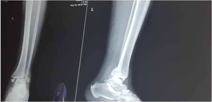
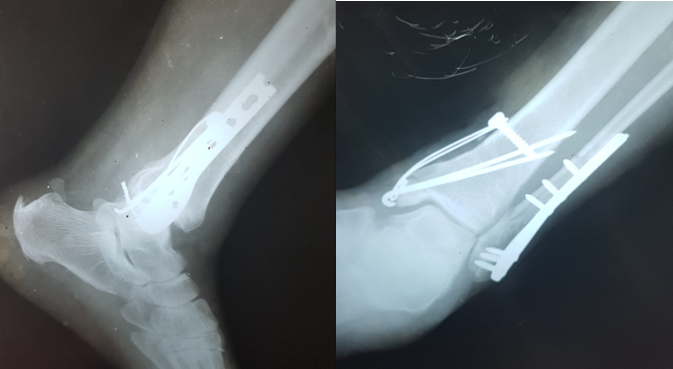
Case 1
40/m closed # bimallolus RT side without DNVD came to Civil Hospital on 17th September 2019 and operated on 18th September 2019 and follows.
Case 2
47/M closed# bimallolus Lt side without DNVD came to Civil Hospital, Ahmedabad on 23th august 2019 and operated on 24th August 2020 and follows.
Conclusion
This prospective, randomised study to compare the result of intramedullary nailing with plating in distal fibula fracture(Weber type B). Anatomical reduction is always important in all intra articular fractures, more so if a weight bearing joint like ankle joint is involved. The fibular length has to be maintained for lateral stability of the ankle.12, 13 The type and extent of injury in ankle fractures is important for good reduction and fixation with good functional outcomes. The aim of surgery should be to achieve anatomical reduction of the fracture fragments, ankle mortise congruity, restoration of the length of the fibula and restoration of syndesmotic integrity.9, 2 During surgery, the soft tissues dissection should be kept minimal to avoid further vascular complications. In the post-operative period, splintage of the ankle should be done and precautions are taken to prevent swelling of the ankle joint as swelling may lead to delayed wound healing.6 Patients are ambulated with crutches or walker without bearing weight on the injured limb from the first post-operative day if there are no associated injuries and can be discharged from the hospital by the first week.7
The four to six week period of immobilization did not affect the final range of ankle function as most patients had achieved full range of motion by the end of 12 weeks postoperatively with active exercise regimen.14
We concluded that fibula plating is a better method of fixation in weber type B and C fractures while intramedullary nailing in fibula is a better method of fixation in weber type A fractures with respect to clinical and functional outcomes.
We also concluded that if the ligament injury has been dealt with properly and repaired and the fixation is anatomically sound, then the period of immobilization (4 to 6 weeks) does not affect the range of motion of ankle joint in the long duration.14


