Introduction
AK Gupta et al. in a clinical study of 32 patients 14 to 62 years reported union in 89.5%. The results were excellent in 15 cases, good in 4 cases and poor in 6 cases. Ununited fracture neck of femur in children and adolescent is a common problem in developing countries where treatment is often delayed. It is a challenging entity to address. The problem associated with delayed presentation are structural bone gap at the fracture site, poor osteogenic potential at fracture margins, hypovascularity of proximal fragment and interposed avascular scar tissue in between the fragment. Osteogenesis or osteosynthesis is the only option in this age group. Different treatment modalities available are open reduction and internal fixation with fibular grafting, valgus osteotomy with and without internal fixation and muscle pedicle bone graft with internal fixation. Although open reduction and internal fixation with or without bone grafting and valgus osteotomy do give acceptable result, the muscle pedicle bone graft has definitely an edge over them as it simultaneously addresses all the problems associated with ununited fracture neck of femur without disturbing proximal femoral anatomy.1 It bridges the structural bone gap, stimulates osteogenesis and improves vascularity of femoral head.2 The aim of anatomical reduction, stable fixation and augmented osteogenesis at fracture site is completely acheived by quadratus femoris muscle pedicle bone grafting with internal fixation resulting in accelerated union and better functional outcome.
Aims of the Study
Present study was undertaken to analyse the result of muscle pedicle bone graft in ununited fracture neck of femur in children and adolescents.
Material and Methods
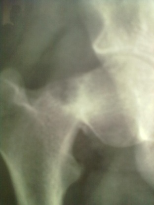
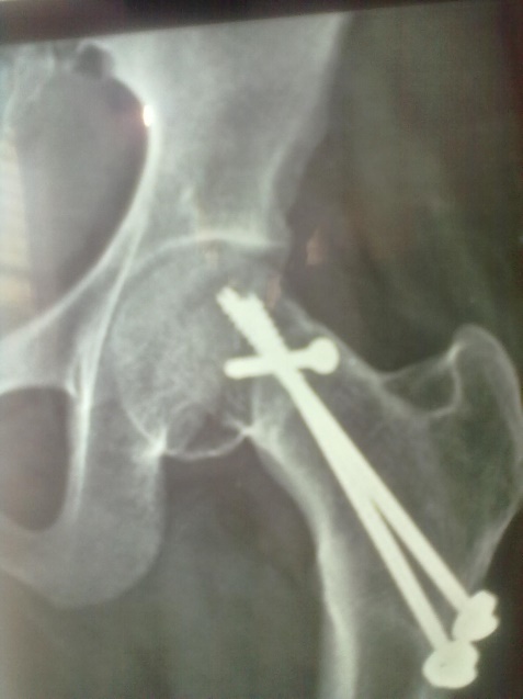
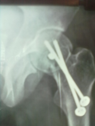
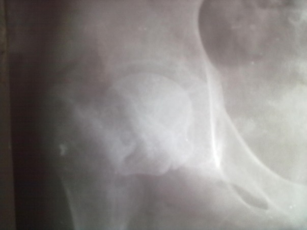
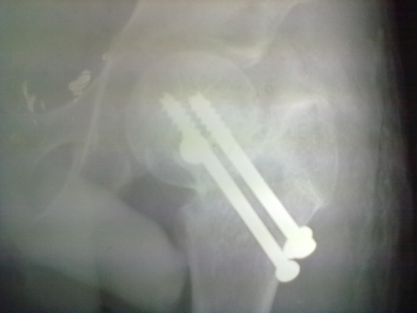
Sixteen patients of ununited fracture neck of femur (presenting late i.e. more than 3 weeks) in the children and adolescent were treated with quadratus femoris based muscle pedicle bone graft with internal fixation between 2004 to 2019. Pathological fracture, recent fracture and compound fracture were excluded from study. Mean age was 14.25 years (standard deviation 1.27 and range 12 to 16 years). Boys dominated our series (n=10 i.e. 62.50%) and right side was more commonly involved (n=12 i.e.75%). All of them were investigated for their fitness for spinal block or general anaesthesia. Steps of procedure including patient positioning, surgical procedure, provisional fixation and definitive fixation were discussed and documented. Radiographic evaluation included A-P View in 15 degree internal rotation, and Lateral view. Average delay in presentation was 5.88 weeks (range was 3-10 weeks). We had 02 transepiphyseal, 11 transcervical and 3 cervicotrochanteric fracture in our series. All the fractures were displaced and having some absorption of femoral neck.
Surgical Procedure
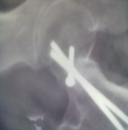
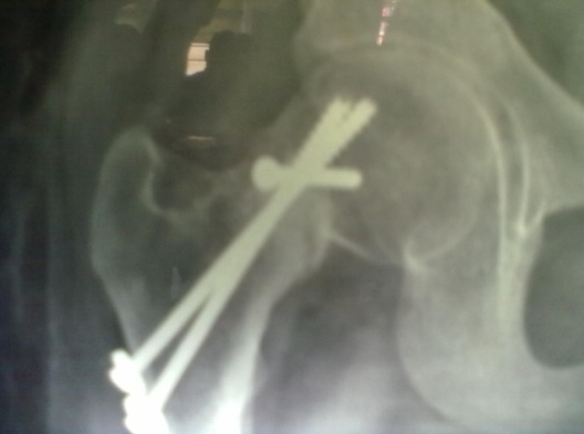
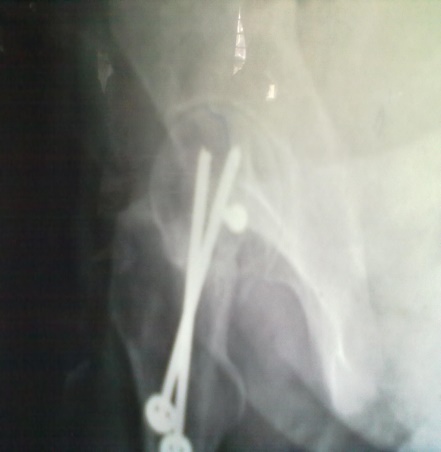
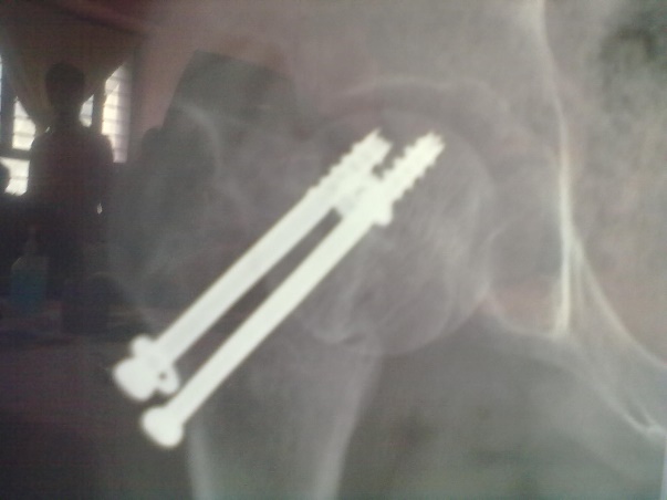
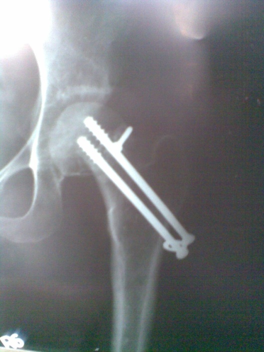
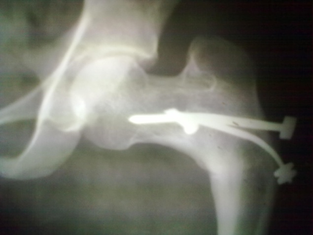
Spinal block was used in the procedure. Prone position on fracture table with radiolucent top was used. Fracture site was exposed through posterior approach and sclerosed margins of the fragments were freshened. Fracture was reduced with appropriate neck shaft angle and fixed with Moors pin/ knowels pin or 4 mm /7mm partially threaded cancellous screws. Any rotation or tilt was corrected. The fixation was augmented with quadratus femoris muscle pedicle bone graft harvested from intertochateric crest area with a bone pedicle of length 2 cm, width 1 cm and depth 1 cm. The graft was secured to the proximal femoral head fragment with 3.5mm cortical screw or 4mm cannulated cancellous screw. Soft tissue closure was done over drain.
Postoperative Care
First dressing change was done on third postoperative day and drain was removed at the same time. Sutures were removed on 14th day. Usually on 5th postoperative day quadriceps exercises was started. Non weight bearing was ensured for 4 months or till radiological union.
Followup
Patients were followed up at 2 weeks, 4 weeks, 6 weeks, 8weeksk, regularly at 6 week interval for next 6 month and then at three month interval up to two year. Radiographic analysis was performed at each follow up with special attention to extent of callus formation, alignment of fragments and hard ware integrity.
Table 1
Shows age distribution of the patient
|
Age of patient (year) |
No. of patient (%) |
|
12 -13 years |
03 (18.75) |
|
13.1-14 years |
04 (25.00) |
|
14.1-15 years |
02 (12.50) |
|
15.1-16 years |
07(43.75) |
Table 2
Shows radiological classification
|
Radiological type |
No. of patient (%) |
|
Transepiphyseal |
02(12.5) |
|
Transcervical |
11(68.75) |
|
Cervicotrochanteric |
03(18.75) |
Table 3
Shows delay in presentatiom
|
Delay (weeks) |
No. of patient (%) |
|
3 |
02(12.50) |
|
4 |
02(12.50) |
|
5 |
04(25.00) |
|
6 |
04(25.00) |
|
7 |
02(12.50) |
|
8 |
01(06.25) |
|
9 |
01(06.25) |
Observation
The result was analysed using Harris hip score and graded as excellent, good, fair and poor.
Discussion
Delayed presntation of fracture neck of Femur in paedriatic age group is not uncommon in developing countries. Meyers Muscle pedicle bone graft has been a well recommended procedure for fracture neck non- union in adult with union rate of 95%.3 Subtrochanteric valgus osteotomy in paedriatic age groups give variable result. 4
Delima DF et al., in a clinical study of 16 patients with ununited transcervical femoral fractures, age 12 to 40 years treated with open reduction and internal fixation with Meyers muscle pedicle graft reported radiological union in 13 patients.5
AK Gupta et al in a clinical study of 32 patients aged 14 to 62 years reported union in 89.5%. The results were excellent in 15 cases, good in 4 cases and poor in 6 cases. Complications like Avascular necrosis (n=2), transient foot drop (n=2), coxa vara (n=1) were noted in the study.2
Harun Yasin Tuzun et al. in his series of 16 patients has reported radiological union at average of 7 months and only one patient (6%) had avascular necrosis.6
In our series of sixteen cases we had 68.75% (n=11) excellent, 12.50% (n=2) good, 12.50 (n=2) fair and 06.25% (n=1) poor result.
Conclusion
Muscle pedicle bone graft with internal fixation has proved to be an efficient method of treatment of ununited fracture neck of femur in children and adolescent as it improves the vascularity of femoral head, bridges the structural gap at fracture site and enhances osteosynthesis at fracture site simultaneously without altering the anatomy of proximal femur.

