Introduction
Intertrochanteric fractures are the commonest fracture encountered in orthopaedic practice. They may be due to trivial domestic fall in elderly osteoporotic bones or road traffic accidents in younger age groups and has significant impact on activities of daily living of the patient.1, 2, 3 Literature shows 5% mortality rate at end of one month and 15% at end of six months after surgery. So the prime aim of treatment is fixation of trochanteric fracture and make the patients pain free and ambulate at the earliest possible and avoiding dependency of bed ridden problems.
The intertrochanteric fractures can be stabilized and internally fixed with different implants right from dynamic hip screw, trochanteric fixation nail, proximal femur nail, Gamma nail, intertan nail, ZNN, PFNa and PFNa2 and surface implants like proximal femur locking plate, Angled blade plate, 95* dynamic hip screw4 and many other newer implants. We all know intramedullary implants scores over surface implants in such fractures. Intramedullary implant has better biomechanical properties and more resistant to failure.5
The author has designed a innovative type of cephalomedullary nail to fix these intertrochanteric fractures which is modification of hip fracture nails available in market. Many of these PFNa, PFNa2 Hip fracture nails have headless cervical screws for fixing head and neck part of femur. All the more the screws penetrate in as you tighten, and whether the compression achieved by these screws on jig is maintained or not is a big question. Secondly the different nails company claim, so and so much amount of compression can be achieved . If more compression is required they cannot achieve the compression and if less compression is required the head screw nail assembly becomes unstable fixation. Finally author has designed a Trochanteric buttress plate along with this nail useful in lateral wall comminution. No other proximal femur implant has Trochanteric buttress plate.
Aims and Objective
A prospective study was done using this single screw innovative nail along with trochanteric buttress plate for fixing the stable as well as unstable variety of intertrochanteric fractures to know its efficacy of fixation, union time and clinical outcome.
Materials and Methods
A study was carried out in Ashwini Sahakari Rugnalaya and Medical research centre Solapur Maharashtra state India from January 2020 to May 2022. A total of 43 cases were operated and followed up and assessed clinically and radiologically every six weeks interval. The age sex and classification of fracture pattern is given in below tabulated format.
Table 4
Classification wise
|
Fracture Type |
Stable /Unstable |
Number of cases |
|
4 Parts |
Unstable |
19 |
|
3 Parts |
Unstable |
13 |
|
2 Parts |
Stable |
7 |
|
IT Basicervical |
Stable |
2 |
|
High Subtrochantric |
|
2 |
|
Total |
|
43 Cases |
Description of Hip fixation nail
Hip Fixation Nail also called as Intra Medullary Hip Screw Nail. It is a cephalomedullary nail with single screw system for fixing intertrochanteric fractures.
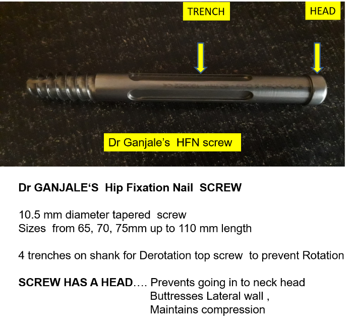
Hip fixation screw has 10.5 mm diameter tapered. It has four trenches on its shank which engages top nut (antirotation screw) from proximal tip of nail. This top nut prevents rotation and allows gradual sliding of cervical screw. The screw has head 12 mm diameter for screwing in to achieve compression. The screw head also has buttressing effect on lateral wall maintaining the constant compression. The screws are available from 65 mm to 110 mm in length and 5 mm difference. Fig 1
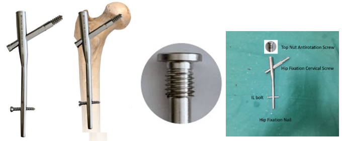
Hip fixation Nail Description: Nail comes with length of 18 cms and Available in diameters ranging from 9 mm to 12 mm diameter and 18 cms long. It has a oblique hole for cervical screw at 130* angle. It has a facility for dynamic interlocking distally with 4.9 mm interlocking bolt. The proximal diameter of nail is 15 mm and has a mediolateral angle of 5 degrees. It is suitable for our Asian especially Indian population who are short statured and smaller femur bone size. (Figure 2)
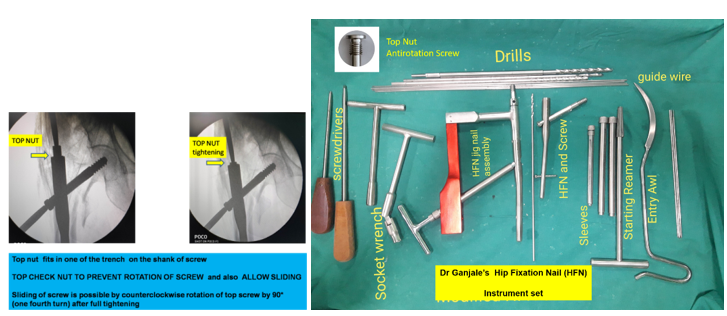
Top Nut /Antirotation screw: A small top nut is screwed in from proximal end of nail which goes in and engages one of the trench on screw shank thus preventing rotation. Figure 2 and Figure 3. This top nut is unscrewed one fourth turn for dynamic fixation to allow gradual sliding of screw enhanching union.
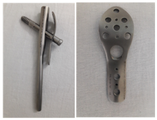
Trochanteric Buttress plate: Author has designed trochanteric Buttress plate along with HFN in case of Lateral wall comminution of greater trochanter. (Figure 4)
Operative procedure
Operative technic is similar to all cephalomedullary nailing procedures. All patients were operated under regional anaesthesia like spinal or epidural anaesthesia. Patient is taken on fracture table with foot attached to foot plates. The normal leg is abducted and flexed to facilitate C- arm movements to view AP and lateral views. Operative leg is adducted for facilitating entry in proximal femur. Painting draping is done. Primary screening of fracture is done in AP and Lateral views in C arm to see the reduction and alignment of fracture. Fracture reduction is held with temperory k wire fixation done anteriorly in lateral view in normal position and then adducted. A 2 cms Skin incision is made 1 to 1.5 inches proximal to Greater trochanter as seen in AP view. A sharp long curved awl is used to have an entry Entry point is just medial to greater trochanter. Awl is passed till lesser trochanter level to open up proximal canal and its placement is checked under c arm. Then a 3 mm guide wire held with T handle chuck is passed through the entry hole into femur passing in distal canal. Now starting reamer is used to enlarge the entry hole in proximal femur over guide wire. Now the HFN Hip Fixation nail is assembled over its jig is passed over guide wire under c arm. The nail placement is monitored under c arm such that its cervical screw hole is aligning at neck head level as confirmed in c arm. Guide wire is drilled through proper sleeve over jig into neck of femur. The placement of guide wire is checked under c arm. It should be in center center in AP and Lateral views of c arm. A inferior placement in AP view and posterior placement in lateral view is accepted to avoid cut out of screw . Now first drill 5 mm cannulated drill is made over properly place guide wire upto subchondral area of head of femur. Later second larger tapered drill is used to drill the neck upto subchondral region in head of femur. A tap 10.5 mm is used to tap the neck for cervical screw. The length of screw is measured from guide wire remaining outside lateral wall of greater trochanter and subtracting the length from second same length guide wire. Hip fixation screw of appropriate length is selected and screwed over pre tapped channel into neck checking under c arm. As we screw the cervical screw into neck we can see compression at fracture site under carm with foot plate traction released, keeping TAD Tip Apex distance 5-10 mm. Distal dynamic interlocking is done through another sleeve over jig. A 4.9 mm Interlocking bolt of appropriate size is locked after drilling a hole. Later jig is removed and a top nut screw is fixed at proximal end of nail. It is also called as antirotation screw. As we tighten the top nut screw it goes in to accommodate in the trench on the shank of cervical screw. This is confirmed by surgeon with respective screw driver in top nut screw and cervical screw and make some finer rotation of cervical screw such that top nut engages in trench and then the cervical screw does not rotate after tightening the top nut screw. The top nut screw is loosened by one fourth turn to allow sliding of cervical screw and enhance fracture union. Final screening of fixation is checked in C arm for confirming the placement of nail cervical screw in head neck and interlocking bolt. Wound wash was given with normal saline and closed in layers. A pressure bandage is applied to operated lower limb thigh.
Trochanteric Buttress plate was used in cases which had comminution of lateral wall. The Trochanteric buttress plate is slided through the incision taken for passing cervical screw, the lateral wall is gently cleared of soft tissue and plate is slided. The hole in plate is aligned with the trajectory for cervical screw and guide wire is passed through hole of plate and into neck portion. Subsequent first drill second drill are used and cervical screw is passed and tightened. Thus, the plate acts as a buttress with the tightened cervical screw. The plate has additional holes for fixing big fragment of greater trochanter. And distally to shaft portion may be unicortical or missing the nail if proximal shaft is wide enough. (Figure 4)
Post operative protocol
Intravenous fluids in post operative period for 12 hours and Antibiotics for three days and later with analgesics A pillow is placed beneath the knee of operated limb for comfort of patient and avoid external rotation of leg. Patient is made to sit on second post operative day Quadriceps exercises are taught and motivated to perform. Patients with stable type of fractures fixed were motivated for partial to full weight bearing walking with walker. Patients with unstable type of fractures is made to ambulate on walker with non weight bearing as per tolerance with supervision. Dressings were done on 3rd, 6th and 10th post operative period Suture removal was done on 12th day. Post operatively. Patient was discharged on 5th post operative day and followed up later at 6 weekly interval for clinical and radiological evaluation.
Results
43 cases were operated with Hip Fixation Nail alone, and 5 cases needed Trochanteric Buttress plate for lateral wall comminution along with Hip fixation nail. All patients were informed about the study in all aspects and an informed consent was obtained. Method of Collection of Data was By interview, clinical assessment using the Kyle’s criteria and radiological examination, By analysing case papers, follow up intervals at every six weeks. The results were Excellent in 31 cases, Good in 5 cases and fair in 2 cases as seen in our study according to Kyles criteria. 5 cases did not follow up after discharge.
Complications
Inadequate reduction due to comminution, subsequent minimal varus of neck as seen in follow up radiological examination but fracture united. No infection was seen in any case. Shortening less than 2 cms as seen in four part unstable fractures but not limiting daily activities.
Follow up:
Highest Follow up 1 yr 3 months
Lowest follow up Immed PO
Expired 5 cases did not turn up for follow up.
Clinical function: we used Kyle’s criteria6
Table 5
KYLE’S criteria
Stable trochanteric fracture cases followed up full weight bearing walking with walker, or walking stick or cane and were most comfortable at six weeks
Unstable trochanteric fracture cases came non weight bearing with walker at 6 weeks and later weaned off the support and started PWB walking at 8 to 10 weeks
Radiological assessment AP and frog leg view was done every six weeks checking for signs of union implant back out . No patients had back out of screw loosening or any infection. 4 part unstable fractures took 10 to 12 weeks to consolidate radiologically.
Discussion
Intertrochanteric fractures can be well managed by Extra medullary surface implants as well as Intra medullary implants. We all know intramedullary implants scores over extramedullary surface implants. Intramedullary implants are very well treated by two screw system like PFN 25 cms length, TFN 18 cms length, Intertan nail for all stable as well as unstable intertrochanteric fractures. Literature mentions single screw system nails like PFNA and PFNA2 helical blade and nail, Gamma Nail, Halifax nail, Hip Fracture Nail, ZNN screw nail and are commonly used for fixing stable and unstable fractures. There are different nail and cannulated screw systems for cement augmented fixation.
Earlier implants like Dynamic hip screw worked on principle of controlled collapse. These were extramedullary implants which had high failure rates in lateral wall fractures and reverse oblique fracture pattern.7, 8, 9 Intramedullary implants proved to have biomechanical advantages.10, 5, 11 Because cephalomedullary nailing (CMN) has shown a clear advantage over the compression hip screw, the indications for CMN have been broadened greatly.12, 13, 14 Tese expanded indications almost all peritrochanteric fractures, including basicervical fracture patterns.
Single screw system like PFNA and PFNa2 are having helical blade to be gently hammered in neck and head. The helical blade in PFNA2 has 2 advantages. It compacts the already weak cancellous bone from the femoral head rather than being removed which happens in femoral screws. It also has more contact surface area with the femoral cancellous bone, than conventional screws2, 15 Bajpai in his study of 77 cases had found that both implants (PFN- screw vs helical) were similar with respect to time of surgery, functional assessment, duration of hospitalization and blood loss.16 At this point the author wants to highlight that drilling with power drills causes much bone loss especially in osteoporotic bones. Overdrilling is another cause in normal bones too, making fixation of helical blade or screw loose, or even exchanging helical blades or screws multiple times in attempt of selecting proper length of helical blade or screw . In screw system one has to be gently ream or drill manually with proper instruments, thus decreasing bone loss and achieving good bony purchase and fixation.
Coming to single screw system available in market, the cervical screw for head neck are smooth and is not having head, The results of single screw of Hip Fracture nail (HFN) screw system of imported companies were excellent and encouraging. These are modified versions of Gama nail.12
The Gamma nail theoretically combines the advantages of intramedullary fixation—lesser surgical trauma and greater mechanical resistance17, 5, 4 with the advantages of the sliding lag screw, which allows controlled fracture collapse. In biomechanical studies, the Gamma nail appeared to be stronger and to reduce the risk of lag screw cutting out.10
What are the problems with. Hip Fracture nail. Many of the Hip Fracture nail screw system available have HEADLESS screws there are possibility of screw penetrating in through nail as we tighten the screw. (Figure 11)
Main question
Whether the compression achieved by the screw with jig nail assembly is maintained in hip fracture nail is questionable ? As you compress the screw by rotation The Sleeve acts as counter against Lateral wall of Trochanter to achieve compression but…… When we disassemble jig the compression achieved on jig is maintained or not is questionable.
TFN, PFN, PFNA, GAMA NAIL, INTERTAN all have different modes of compression
TFN PFN achieves compression by partially threaded lag screws,
To achieve compression They depend on 2 things
So whenever these two things are compromised we cannot get good compression, Secondly we have to use Trochanteric Buttress Plate supports screw heads preventing varus drift. One can achieve compression by utilising nail surface as lateral wall and get compression by tightening screw over that surface. This is possible in younger patients with good bone stock. In osteoporotic bones the lateral wall is so thin it gets crushed with tightening the screws making point of contact on lateral wall loosing its stability and vulnerable to go into varus and cut out later.
In single screw system Hip Fracture nail the screw has to be placed just outside the bone surface for gradual sliding and enhance healing. If kept flush to bone new bone growth occurs over screw tip distally at trochanter, the sliding of cervical screw is prevented in due course and removal becomes difficult and we will have to dig out lateral wall for retrieval of screw.5
Cost of implant of Hip Fracture nail is not affordable and not easily available at smaller places away from city.
Considering above drawbacks the author has designed and Modified Hip Fracture nail and named as Hip Fixation Nail with single Screw system, also called as Intramedullary Hip Screw Nail IMHSN. It is almost similar to Hip Fracture nail, Gama nails and ZNN nail single screw system but with a difference mentioned in description of nail and screw.
Three point proximal femur fixation depends on good subchondral purchase of screw in head, Lateral wall intactness and the Greater trochanter entry point of any cephalomedullary nail. Two important points as circled are predictors of failure. Surgeon has 2 points in his control a good subchondral fixation of screw with perfect tip apex distance TAD, (within 10 to 15 mm) and lateral wall comminution can be overcome by buttressing effect with trochanteric buttress plate. The comminuted Greater trochanter is the factor not in surgeons control. This study has demonstrated that mechanical failure of the Gamma nail in peri-trochanteric femoral fractures is rare (< 1%) when three-point proximal fixation is achieved.8 However, it is also noted a high failure rate when the lateral end of the lag screw (which is headless) was positioned short of the lateral femoral cortex.5
In most prior studies, a standard Gamma nail device has been used, having a length of 200 mm, mediolateral curvature of 10*, proximal diameter of 17 mm, and available distal diameters between 12 and 16 mm, but some other authors have used variations of the nail design. Aune et al used a modified nail with a medio-lateral curvature of 6* and distal diameters between 12 and 14 mm, and they reported 10 out of 177 intraoperative femoral shaft fractures, 7 of which had distal locking with 2 screws. Leung et al used a modified nail for the Asian anthropometry that had a length of 180 mm, mediolateral curvature of 4*, proximal diameter of 16 mm, and distal diameters of 11 and 12 mm; they reported 4 out of 349 intraoperative fractures of the greater trochanter but no cases of postoperative femoral shaft fractures. This modified design of Gamma nail was associated with a lower rate of postoperative complications than with the standard Gamma nail.18
Considering above points, the screw designed by author has head 12 mm diameter which buttresses and remains outside on lateral wall with good subchondral purchase in head of femur and fulfills the factors for better fixation. As the screw is tightened there is compression at fracture site and is not dependent on any sleeve as it occurs in Hip Fracture nail, gama, ZNN nail screw system. The amount of compression required is independent and is not limited according to company set up.
Anatomical variations of proximal femur in shape and size of neck especially short statured asian community and Indians females are commonly encountered. These patients have short neck and diameter so, two screw system nail is difficult to accommodate in neck. This single screw system nail is very useful in such cases. Secondly there is lot of area in neck to err for placing 10.5 mm screw in the narrow neck. The proximal diameter of author’s nail is 15 mm much smaller than 17mm as seen in gama nails especially useful for tall built and thicker proximal femur. The mediolateral angle is 5 * which is easier to pass just medial to greater trochanter into proximal shaft of femur.
Conclusion
Hip Fixation Nail designed by author can be used to fix all stable as well as unstable intertrochanteric fractures. There is much space is available in neck to err for placing guide wire and screw. It has excellent stable fixation with antirotation top screw. It allows sliding collapse for union. Very useful in elderly osteoporotic hips. Gentle screwing achieves compression of fracture site. Head of screw acts as buttress on lateral wall.
Trochanteric buttress plate is available in case of lateral wall comminution which is not available in any other single screw system.
Simple instrumentation easy to use-surgeon friendly. It is very much cost effective and cheaper than imported nails and is easily available with excellent results and functional outcome. Despite of lot of comparative studies there is no clear cut guidelines regarding to which implant should be used and when. The main factors deciding the final outcome of trochanteric fractures fixation depend on fracture geometry, bone quality, age, type of fixation, placement of implant, technic of fixation, surgical skill and experience of surgeon.


