Introduction
Kuntscher cloverleaf nail, also called as K-nail, is an intramedullary nail (IMN), made from stainless steel with a slot along the longitudinal axis. It is used in fixation of short oblique, simple transverse, Winquist- I and II comminuted fractures of femur.1 3-point fixation using metal IMNs attached to endosteal surface and the concept of elastic nailing was 1st identified by Gerhard Kuntscher et al.2, 3 at the university of Kiel Germany during 1930s. Initially, the nail was in shape of “V” but later, it was introduced in four-leaved clover form for more strength and easy usage. K nail migration inside medullary canal of femur is one of the common complications. But spontaneous migration of K-nail distally beyond tibial tuberosity or bypassing the bone along soft tissue of knee and leg is a complication that was rarely seen. Distal migration of K-nail was 1st reported during 1942s. Common reasons for extrusion include delayed union, infection, incorrect K-nail size, osteoporosis, osteogenesis imperfecta, performing fixation by faulty methods, pseudoarthrosis, after surgery in association with oblique fractures of lower 1/3rd of shaft of femur and premature weight bearing.4, 5, 6, 7 It is suggested that K-nails to be extracted immediately after union and radiological consolidation of fracture. This was a case in which the Kuntscher nail was migrated distally, most probably perforating distal femoral surface through the oblique fracture line and slowly migrating across knee joint from the posterior aspect and entering the posterior compartment of leg till the ankle. In this case, we suspected the migration of K-nail due to malunion of an oblique distal femur fracture with anterior displacement. There were no recent reports on this type of complete extrusion.
Case Report
A 72-year-old male, who is working as a farmer in a rural area, came to our institution with pain and swelling on & off in the left ankle for last two months during May 2022. History revealed that he developed pain in the left ankle which was insidious in onset, continuous in nature, aggravated by walking and relieved with rest. He experienced swelling in the left ankle on walking which used to get relieved with rest and elevation. He sustained a closed fracture in the lower 1/3rd of left femur due to fall from a tree 25 years ago for which he underwent Kuntscher nailing and those records were not available. Postoperative period was uneventful and patient strictly followed non-weight bearing for 3 months after surgery. Patient was asymptomatic for 25 years and was able to do his daily activities. He experienced discomfort in the left knee joint during flexion, disabling him to squat and sit cross-legged for past two months. He had no comorbidities.
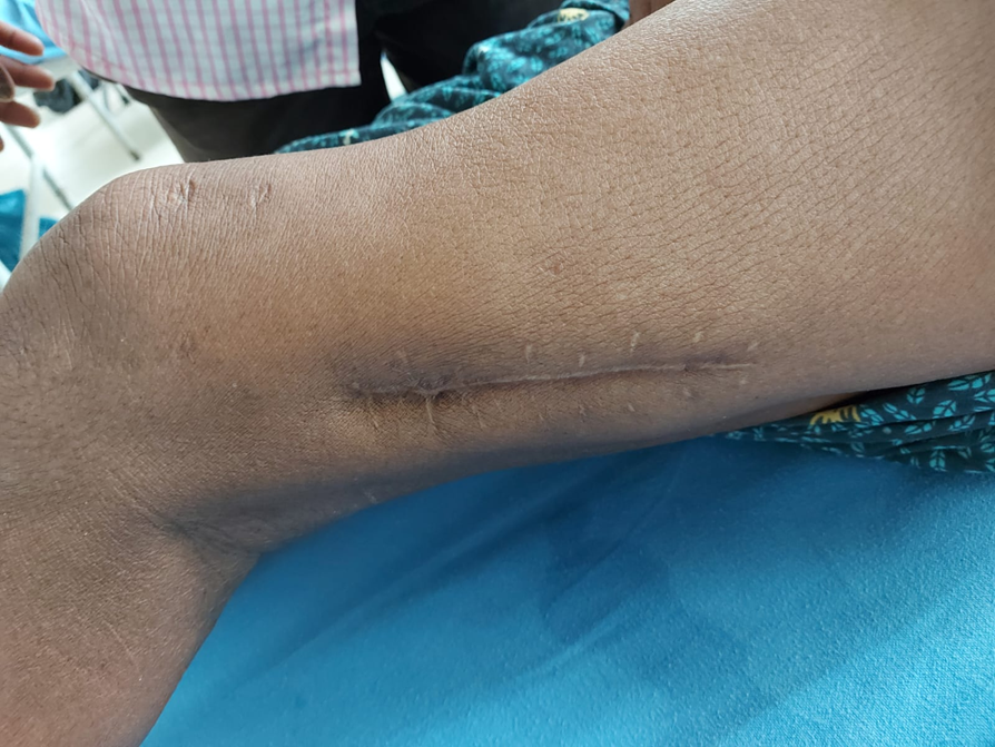
Local examination of left lower limb revealed swelling around the left ankle. There was no redness or sinuses or engorged veins or visible pulsations. There is a single healed surgical scar of length 10 centimeters on the lateral aspect of left distal thigh.
On palpation, there was non-pitting edema around the left ankle. There was no local rise of temperature or tenderness. There was a hard, metallic implant palpable just posterior to the left medial malleolus. There were no other palpable swellings or lymph nodes and no distal neurovascular deficit.
All the ankle joint movements were normal and painless. But the flexion of left knee was restricted beyond 90o and caused slight discomfort to the patient. There was no limb length discrepancy.
We suggested for, X-ray left thigh with hip and knee in AP and lateral view, and X-ray left leg with knee & ankle in AP & Lateral views.
We finally diagnosed the case as – Distal migration of Kuntscher nail into leg used for treatment of 25 years old malunited fracture distal 1/3rd of left femur and planned for implant removal.
Surgery
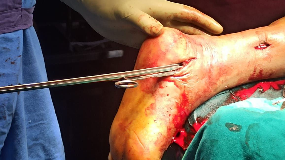
A 5 cm incision was given inferior and posterior to left medial malleolus, subcutaneous planes dissected and tip of the K-nail was identified. All the adhesions and fibrous tract around the nail was cleared and K-nail extracted. The patient was discharged in a stable condition and 6 months follow-up gave a good functional outcome.
Discussion
In the previous era where treatment of femur fracture was just traction or cast splintage, K-nailing has become the main mode or gold standard mode of management. But there are various pitfalls of K nail, like collapse, failure to prevent migration, rotation of fragments of fracture, especially in inherently unstable fractures. With the introduction of “locking” using bolts at each end of the modern nail, open K-nails are not suggested for fixation of femoral shaft fractures. Cause for distal migration in our patient could be due to malunion of a long oblique fracture with anterior displacement or faulty fixation. Our patient was a 72-year-old male, who presented 25 years later with migration of K-nail into the posterior compartment of leg which was fixed for distal femur fracture, without any neurovascular injury. It was surprising in our case to see no significant complaints apart from pain and swelling over the ankle. One author reported a case 12 years postinsertion in a 33-year-old female who came with a complaint of sudden onset of left knee pain and examination showed extrusion of Kuntscher nail 12 years postinsertion. She underwent K-nail removal and showed better outcomes after 6 months of follow-up. Cases reported by De Belder et al.8 showed fractures in proximal half that involved isthmus; and only 1 had lower most femoral fracture. Previous studies done doesn’t cover the information on the fracture configuration which is more prone to migration.1, 9
Siddharth Jain et al.10 reported migration of k nail in a 41-year-old man who presented with a sinus which is discharging pus over tibial tuberosity since 12 months and K-nail was kept six years back.
Presence of K-nail for long time (25 years) may have also produced various components of ionization that accumulated between the nail and the bone and might have caused expulsion of the nail. Before introduction of K-nails by Gerhard Kuntscher management of femur fractures was restricted to traction or cast splintage, which required more duration of inactivity. Kuntscher nailing caused early return to activity, mostly with in few weeks, as the nails share load with the bone, instead of entirely supporting the bone. But pitfalls of K nails which include migration or collapse can be addressed by "locking" of modern IM nails with the help of bolts at each end of nail, fixing the nail to bony cortex. From the introduction of closed interlocking nails of the femur, open K-nails are not routinely used for fixation of femoral shaft fractures.11, 12 But it is still indicated in hospitals where image intensifier or traction tables are not available and in cases where the patient can’t afford expenses of interlocking nails, which is the scenario in most of the developing countries. Sometimes it was not possible to assess the cause of migration of K-nail using imaging features or any others investigations. Loose fitting nails causing continuous movement at fracture site can cause migration.
Conclusion
Distal migration of K-nail across the knee joint and into the leg after femur diaphyseal fracture fixation is a rare and preventable complication. It is recommended that K-nails should be routinely removed as soon as radiological union and consolidation of the fracture is confirmed, to avoid rare postoperative complication of distal migration of the K-nail.


