- Visibility 78 Views
- Downloads 4 Downloads
- DOI 10.18231/j.ijos.2024.006
-
CrossMark
- Citation
Comparative analysis of knee radiographs between weight bearing AP and Rosenberg PA flexion view for knee osteoarthrosis
Introduction
Osteoarthritis (OA) of the knee is one of the commonest cause of pain & disability worldwide affecting women predominantly. It is a chronic degenerative disease of varied etiology with an estimated prevalence of about 28.7% in India which increases with age.[1] The disease commonly affects the knee joint and is characterized by the joint space narrowing due to loss of articular cartilage, marginal hypertrophy of the bone also known as osteophytes, subchondral sclerosis and cysts, and inflammation of the synovial membrane and joint capsule; the cartilage ultimately softens, ulcerate & focally disintegrate in the later stages.[2]
Plain radiographs has been the ancient & the most cost effective diagnostic tool for assessing articular cartilage wear & tear in osteoarthritis of the knee & studies have shown to optimize the information obtained from plain radiographs to enhance its sensitivity & decrease the need for alternative imaging studies.[3] Radiographs demonstrate the severity, extent & progression of knee OA by determining the joint space narrowing (JSN) in the tibio-femoral compartment of the knee.[4]
Rosenberg PA flexion view has been shown to be more sensitive to detect JSN in the medial & lateral compartments of the knee as compared to the conventional AP weight bearing view.[5] The JSN helps in the early diagnosis of OA[6], [7] & correlates significantly with the patient’s knee injury and osteoarthritis outcome score (KOOS).[8]
We hypothesized that the combination of the weight bearing AP & Rosenberg PA flexion view can help to detect early JSN in the medial and lateral compartments of the knee as well as detect early degenerative changes thus increasing the radiographic sensitivity of OA. The aim of this study is to evaluate, quantify & compare the two radiographic views to improve early detection of JSN in knee osteoarthritis (OA) using Kellgren-Lawrence classification (KL), Ahlback grading & International Knee Documentation Committee (IKDC).
Materials and Methods
This was a retrospective study involving 31 patients (47 knees) visiting the out patient department of a tertiary care facility in Telangana during the period of Jan 2014 to Dec 2018. All patients above the age of 18 years complaining of knee pain were included in the study. Patients with secondary osteoarthritis of the knee, previous surgeries on the knee, flexion deformity of the knee more than 30 degrees were excluded from the study.
Each patient who underwent assessment after a detailed history and primary examination followed by clinical examination of both the knees, who were subjected to a conventional weight bearing AP view of the knee in full extension and the standing Rosenberg PA flexion view with knee in 45 degrees of flexion were included in the study.[9] The radiographic assessment of Rosenberg view is demonstrated by centered exposure of bilateral knees without rotation. The X-ray tube was centered on the patella and placed approximately 100 cm away directed 10 degrees caudal with both the patellae touching the cassette. Radiographs of both knees were obtained for comparison.[10] The Rosenberg view is not taken at a perpendicular angle hence allows for more visualization of tibia. Diagrammatic representation of the position of Rosenberg view is shown in [Figure 1].

All radiographs were analyzed with their corresponding knee scores. The Kellgren-Lawrence classification, Ahlback grading and IKDC scores were used to grade the severity of the knee osteoarthritis. The IKDC grading of severity A, B, C or D was converted into numerical order of 1, 2, 3 & 4 for the convenience of comparison purposes.
A printed digital radiographs of both the knees without magnification were used to document, quantify and compare the joint space narrowing and osteophytes. A vernier caliper was used to measure the medial and lateral tibio-femoral compartments and compared with the other side.
Results
There were 13 males and 18 females in our study population comprising 47 knees, 25 right & 22 left. Average age of the study population was 53.8 years (range: 45-70). Four patients had a previous history of injury to the knee; six patients had comorbidities like hypertension & diabetes mellitus and 29 out of 31 patients were using drugs for knee pain with Apleys pain grading as grade I in 8.3%, grade II in 55.6%, grade III in 33.3% & grade IV in 2.8%. The medial & lateral joint space measurement showed early and significant joint space narrowing on Rosenberg PA flexion view and had a significantly high correlation with patients clinical as well as radiological scores.
Kellgren-Lawrence (KL) & Ahlback classification were used to score the knee using the radiological data, while the IKDC was used for objective evaluation grading of the knee. The Rosenberg view increased the severity of KL scores in 7 knees; 2 knees changed severity score from previous grade I to grade III, 3 knees from grade II to III and 2 knees from grade III to IV. Similarly, in Ahlback classification the Rosenberg view increased the severity of scores in 12 knees, while it decreased in 1 knee; 1 knee changed severity score from previous grade 0 to grade III while 1 knee changed from previous grade I to grade 0, 2 knees from grade I to II, 6 knees from grade I to III, 2 knees from grade II to III & 1 knee from grade III to IV. The IKDC score was increased in 14 knees & decreased in 3 knees with the Rosenberg view; 2 knees changed the severity of the previous grade II to III, 3 knees decreased from grade IV to III & 12 knees increased from grade III to IV.
On statistical analysis of the 3 groups between the AP & Rosenberg view using paired t test, the difference was statistically significant in all the 3 groups ([Table 1]). Furthermore on cross comparison of the paired groups, the difference was also found to be statistically significant ([Table 2]).
|
Paired t test groups (n=47) |
Mean |
Std. deviation |
Correlation |
Sig. |
|
|
Pair 1 |
KL-AP |
2.72 |
0.826 |
0.806 |
<0.005 |
|
KL-R |
2.91 |
0.747 |
|||
|
Pair 2 |
AB-AP |
2.09 |
0.974 |
0.609 |
<0.005 |
|
AB-R |
2.49 |
0.882 |
|||
|
Pair 3 |
IKDC-AP |
3.38 |
0.644 |
0.582 |
<0.005 |
|
IKDC-R |
3.62 |
0.573 |
|
Wilcoxon Signed Ranks Test |
||
|
n=47 |
Z |
Asymp. Sig. |
|
AB-AP & KL-AP |
-3.985 |
<0.005 |
|
AB-R & KL-R |
-3.386 |
0.001 |
Discussion
Kellgren & Lawrence, Ahlback & the IKDC are a few classifications described for the grading & severity of OA knee based on JSN & degenerative changes.[11], [12], [13]
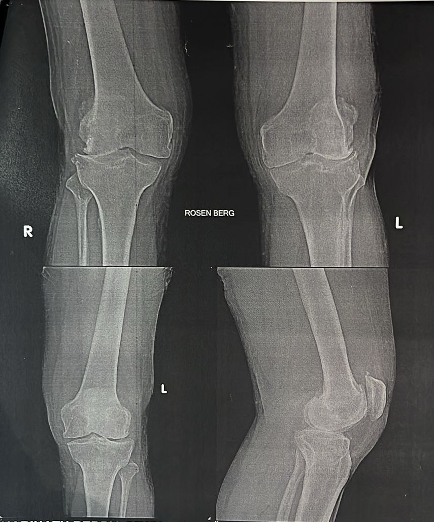
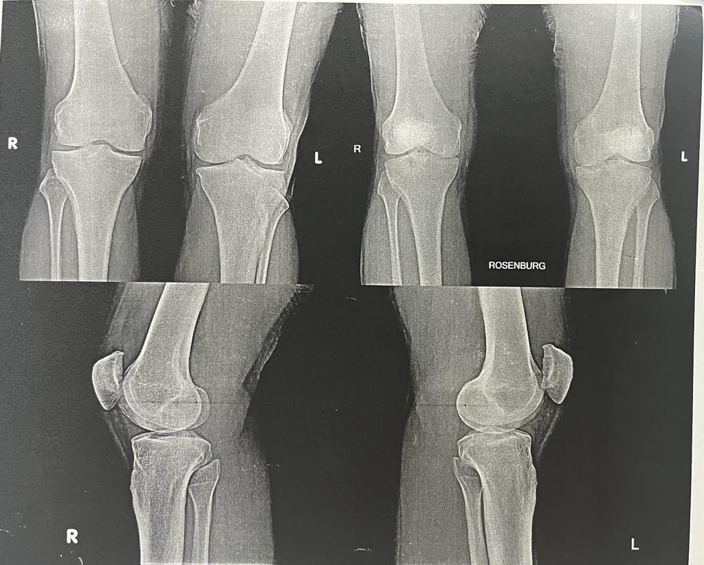
The radiological evaluation of the knee pain most commonly comprises of weight bearing AP, lateral & the skyline radiographs of the knee joint. This study evaluates the advantages of an additional imaging view Rosenberg PA flexion view in early radiological detection of OA. [Figure 2], [Figure 3] are examples suggesting early detection of joint space narrowing in Rosenberg PA view as compared to standard AP view. Many prior studies have compared this flexion view directly to the AP view but none has compared it with 3 different grading classifications in Indian population. Most studies have compared the AP view directly to the flexion view, however in this study we have used both views & compared with 3 different grading systems of knee.
Rosenberg et al. in the study of 55 knees directly compared the AP & flexion view radiographs & concluded that the flexion view correlates better with arthroscopic findings.[9] Merle-Vincent F et al. in a study of 202 knees in 2007 concluded that the standing AP radiograph alone is poor in diagnosing the severity of JSN in early OA.[7] In a study of 50 patients, Ritchie et al demonstrated that the Schuss view radiograph is a valuable tool in early OA which can alter the clinical management,[14] while a prospective analysis by Davies et al. supports the flexion view over the standard extension view.[15] All these studies compared the varying degrees of flexion view of the knee joint with the standard weight bearing view in extension to aid in early diagnosis of OA.
In our study, the Rosenberg view significantly aided in early diagnosis of JSN when compared to the standard weight bearing AP radiographs which is comparable to the study by Yamanaka et al,[16] where early identification of JSN was more significant in medial compartment of the knee as compared to the lateral compartment, possibly because of heavier loading in the medial compartment.[17] The KL, Ahlback & IKDC severity scores increased significantly from the previous grade on Rosenberg view as compared to the standard weight bearing AP radiographs.
In addition to the JSN, the Rosenberg views provided significant identification of subchondral cysts, sclerosis of joint line, osteophytes at the margins, intercondylar notch & the tibial spine. This improved visualization is attributed to the natural tibial slope which gets overlapped with femoral condyles in full extension.


Conclusion
The Rosenberg PA flexion view is an important tool which when used in conjunction with the standard weight bearing AP radiographs of the knee helps in early diagnosis of OA. The KL, Ahlback and IKDC scores are best evaluated earliest on the Rosenberg views.
Limitations
Small sample size is the limitation of our study.
Source of funding
None
Conflict of Interest
None.
References
- CP Pal, P Singh, S Chaturvedi, KK Pruthi, A Vij. Epidemiology of knee osteoarthritis in India and related factors. Indian J Orthop 2016. [Google Scholar]
- DT Felson, TE Mcalindon, JJ Anderson, A Naimark, BW Weissman, P Aliabadi. Defining radiographic osteoarthritis for the whole knee. Osteoarthritis Cartilage 1997. [Google Scholar]
- GF Dervin, RJ Feibel, K Rody, J Grabowski. 3-Foot standing AP versus 45 degrees PA radiograph for osteoarthritis of the knee. Clin J Sport Med 2001. [Google Scholar]
- E Vignon, M Piperno, MPHL Graverand, SA Mazzuca, KD Brandt, P Mathieu. Measurement of radiographic joint space width in the tibiofemoral compartment of the osteoarthritic knee: comparison of standing anteroposterior and Lyon schuss views. Arthritis Rheum 2003. [Google Scholar]
- RC Fontboté, UF Nemtala, OO Contreras, R Guerrero. Rosenberg projection for the radiological diagnosis of knee osteoarthritis. Rev Med Chil 2008. [Google Scholar]
- M Piperno, MPHL Graverand, T Conrozier, M Bochu, P Mathieu, E Vignon. Quantitative evaluation of joint space width in femorotibial osteoarthritis: comparison of three radiographic views. Osteoarthritis Cartilage 1998. [Google Scholar]
- Merle-Vincent F Vignon, E Brandt, K Piperno, M Coury-Lucas, F Conrozier, T. Superiority of the Lyon schuss view over the standing anteroposterior view for detecting joint space narrowing, especially in the lateral tibiofemoral compartment, in early knee osteoarthritis. Ann Rheum Dis 2007. [Google Scholar]
- SR Oak, A Ghodadra, CS Winalski, A Miniaci, MH Jones. Radiographic joint space width is correlated with 4-year clinical outcomes in patients with knee osteoarthritis: data from the osteoarthritis initiative. Osteoarthritis Cartilage 2013. [Google Scholar]
- TD Rosenberg, LE Paulos, RD Parker, DB Coward, SM Scott. The forty-five-degree posteroanterior flexion weight-bearing radiograph of the knee. J Bone Joint Surg Am 1988. [Google Scholar]
- RB Mason, JG Horne. The posteroanterior 45 degrees flexion weight-bearing radiograph of the knee. J Arthroplasty 1995. [Google Scholar]
- JH Kellgren, JS Lawrence. Radiological assessment of osteo-arthrosis. Ann Rheum Dis 1957. [Google Scholar]
- S Ahlbäck. Osteoarthrosis of the knee. A radiographic investigation. Acta Radiol Diagn (Stockh) 1968. [Google Scholar]
- NJ Collins, D Misra, DT Felson, KM Crossley, EM Roos. Measures of knee function: International Knee Documentation Committee (IKDC) Subjective Knee Evaluation Form, Knee Injury and Osteoarthritis Outcome Score (KOOS), Knee Injury and Osteoarthritis Outcome Score Physical Function Short Form (KOOS-PS), Knee Outcome Survey Activities of Daily Living Scale (KOS-ADL), Lysholm Knee Scoring Scale, Oxford Knee Score (OKS), Western Ontario and McMaster Universities Osteoarthritis Index (WOMAC), Activity Rating Scale (ARS), and Tegner Activity Score (TAS). Arthritis Care Res (Hoboken) 2011. [Google Scholar]
- JFS Ritchie, M Al-Sarawan, R Worth, B Conry, PA Gibb. A parallel approach: the impact of schuss radiography of the degenerate knee on clinical management. Knee 2004. [Google Scholar]
- AP Davies, DA Calder, T Marshall, MM Glasgow. Plain radiography in the degenerate knee. A case for change. J Bone Joint Surg Br 1999. [Google Scholar]
- N Yamanaka, T Takahashi, N Ichikawa, H Yamamoto. Posterior-anterior weight-bearing radiograph in 15 degree knee flexion in medial osteoarthritis. Skeletal Radiol 2003. [Google Scholar]
- BL Wise, J Niu, M Yang, NE Lane, W Harvey, DT Felson. Patterns of compartment involvement in tibiofemoral osteoarthritis in men and women and in whites and African Americans. Arthritis Care Res (Hoboken) 2012. [Google Scholar]
